Cryo-electron tomography of Birbeck granules reveals the molecular mechanism of langerin lattice formation.
Oda, T., Yanagisawa, H., Shinmori, H., Ogawa, Y., Kawamura, T.(2022) Elife 11: e79990-e79990
- PubMed: 35758632 Search on PubMedSearch on PubMed Central
- DOI: https://doi.org/10.7554/eLife.79990
- Primary Citation Related Structures:
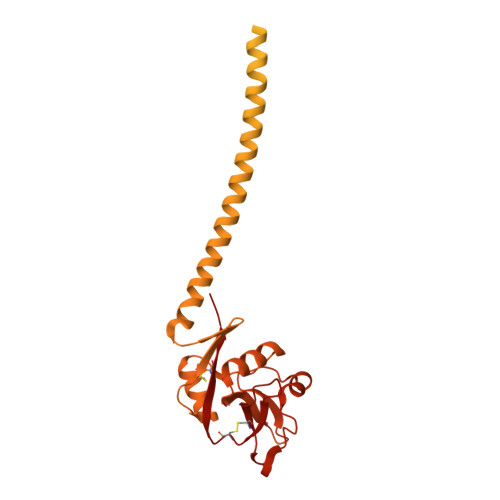
7WZ8 - PubMed Abstract:
Langerhans cells are specialized antigen-presenting cells localized within the epidermis and mucosal epithelium. Upon contact with Langerhans cells, pathogens are captured by the C-type lectin langerin and internalized into a structurally unique vesicle known as a Birbeck granule. Although the immunological role of Langerhans cells and Birbeck granules have been extensively studied, the mechanism by which the characteristic zippered membrane structure of Birbeck granules is formed remains elusive. In this study, we observed isolated Birbeck granules using cryo-electron tomography and reconstructed the 3D structure of the repeating unit of the honeycomb lattice of langerin at 6.4 Å resolution. We found that the interaction between the two langerin trimers was mediated by docking the flexible loop at residues 258-263 into the secondary carbohydrate-binding cleft. Mutations within the loop inhibited Birbeck granule formation and the internalization of HIV pseudovirus. These findings suggest a molecular mechanism for membrane zippering during Birbeck granule biogenesis and provide insight into the role of langerin in the defense against viral infection.
- Department of Anatomy and Structural Biology, Graduate School of Medicine, University of Yamanashi, Chuo, Japan.
Organizational Affiliation: