7WW9

Crystal structure of MutT-8-oxo-dGTP complex: Reaction for 1.5 hr in 20 mM Mn2+


Experimental Data Snapshot

  • Method: X-RAY DIFFRACTION
  • Resolution: 1.80 Å
  • R-Value Free: 0.178 
  • R-Value Work: 0.161 
  • R-Value Observed: 0.162 

wwPDB Validation   3D Report Full Report


Ligand Structure Quality Assessment 


This is version 1.1 of the entry. See complete history


Literature

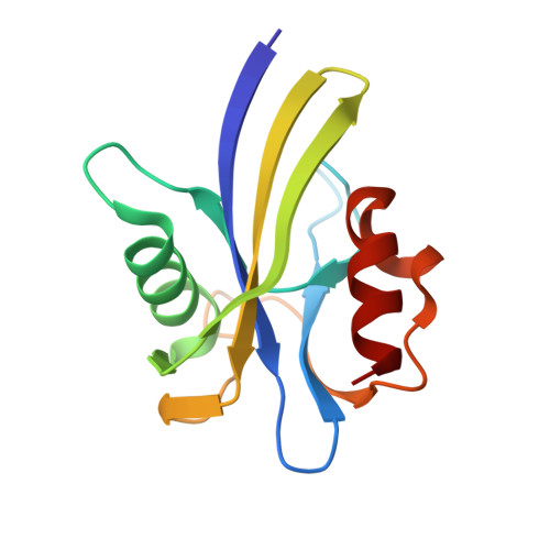
Visualization of mutagenic nucleotide processing by Escherichia coli MutT, a Nudix hydrolase.

Nakamura, T.Yamagata, Y.

(2022) Proc Natl Acad Sci U S A 119: e2203118119-e2203118119

  • DOI: https://doi.org/10.1073/pnas.2203118119
  • Primary Citation of Related Structures:  
    7WW5, 7WW9, 7X9N

  • PubMed Abstract: 

    Escherichia coli MutT prevents mutations by hydrolyzing mutagenic 8-oxo-2'-deoxyguanosine 5'-triphosphate (8-oxo-dGTP) in the presence of Mg2+ or Mn2+ ions. MutT is one of the most studied enzymes in the nucleoside diphosphate-linked moiety X (Nudix) hydrolase superfamily, which is widely distributed in living organisms. However, the catalytic mechanisms of most Nudix hydrolases, including two- or three-metal-ion mechanisms, are still unclear because these mechanisms are proposed using the structures mimicking the reaction states, such as substrate analog complexes. Here, we visualized the hydrolytic reaction process of MutT by time-resolved X-ray crystallography using a biological substrate, 8-oxo-dGTP, and an active metal ion, Mn2+. The reaction was initiated by soaking MutT crystals in a MnCl2 solution and stopped by freezing the crystals at various time points. In total, five types of intermediate structures were refined by investigating the time course of the electron densities in the active site as well as the anomalous signal intensities of Mn2+ ions. The structures and electron densities show that three Mn2+ ions bind to the Nudix motif of MutT and align the substrate 8-oxo-dGTP for catalysis. Accompanied by the coordination of the three Mn2+ ions, a water molecule, bound to a catalytic base, forms a binuclear Mn2+ center for nucleophilic substitution at the β-phosphorus of 8-oxo-dGTP. The reaction condition using Mg2+ also captured a structure in complex with three Mg2+ ions. This study provides the structural details essential for understanding the three-metal-ion mechanism of Nudix hydrolases and proposes that some of the Nudix hydrolases share this mechanism.


  • Organizational Affiliation

    Graduate School of Pharmaceutical Sciences, Kumamoto University, Kumamoto, 862-0973, Japan.


Macromolecules
Find similar proteins by:  (by identity cutoff)  |  3D Structure
Entity ID: 1
MoleculeChains Sequence LengthOrganismDetailsImage
7,8-dihydro-8-oxoguanine-triphosphatase129Escherichia coliMutation(s): 0 
EC: 3.6.1.55 (PDB Primary Data), 3.6.1 (PDB Primary Data)
UniProt
Find proteins for P08337 (Escherichia coli (strain K12))
Explore P08337 
Go to UniProtKB:  P08337
Entity Groups  
Sequence Clusters30% Identity50% Identity70% Identity90% Identity95% Identity100% Identity
UniProt GroupP08337
Sequence Annotations
Expand
  • Reference Sequence
Small Molecules
Ligands 5 Unique
IDChains Name / Formula / InChI Key2D Diagram3D Interactions
8DG (Subject of Investigation/LOI)
Query on 8DG

Download Ideal Coordinates CCD File 
B [auth A]8-OXO-2'-DEOXYGUANOSINE-5'-TRIPHOSPHATE
C10 H16 N5 O14 P3
BUZOGVVQWCXXDP-VPENINKCSA-N
8OG
Query on 8OG

Download Ideal Coordinates CCD File 
C [auth A]8-OXO-2'-DEOXY-GUANOSINE-5'-MONOPHOSPHATE
C10 H14 N5 O8 P
AQIVLFLYHYFRKU-VPENINKCSA-N
SO4
Query on SO4

Download Ideal Coordinates CCD File 
D [auth A]SULFATE ION
O4 S
QAOWNCQODCNURD-UHFFFAOYSA-L
MN
Query on MN

Download Ideal Coordinates CCD File 
E [auth A],
F [auth A],
G [auth A]
MANGANESE (II) ION
Mn
WAEMQWOKJMHJLA-UHFFFAOYSA-N
NA
Query on NA

Download Ideal Coordinates CCD File 
H [auth A]SODIUM ION
Na
FKNQFGJONOIPTF-UHFFFAOYSA-N
Experimental Data & Validation

Experimental Data

  • Method: X-RAY DIFFRACTION
  • Resolution: 1.80 Å
  • R-Value Free: 0.178 
  • R-Value Work: 0.161 
  • R-Value Observed: 0.162 
  • Space Group: P 21 21 21
Unit Cell:
Length ( Å )Angle ( ˚ )
a = 37.962α = 90
b = 55.906β = 90
c = 59.12γ = 90
Software Package:
Software NamePurpose
PHENIXrefinement
HKL-2000data reduction
HKL-2000data scaling

Structure Validation

View Full Validation Report


Ligand Structure Quality Assessment 


Entry History & Funding Information

Deposition Data


Funding OrganizationLocationGrant Number
Ministry of Education, Culture, Sports, Science and Technology (Japan)Japan--

Revision History  (Full details and data files)

  • Version 1.0: 2022-06-01
    Type: Initial release
  • Version 1.1: 2023-11-29
    Changes: Data collection, Refinement description