7WUQ

Tethered peptide activation mechanism of adhesion GPCRs ADGRG2 and ADGRG4


Experimental Data Snapshot

  • Method: ELECTRON MICROSCOPY
  • Resolution: 2.90 Å
  • Aggregation State: PARTICLE 
  • Reconstruction Method: SINGLE PARTICLE 

wwPDB Validation   3D Report Full Report


This is version 1.1 of the entry. See complete history


Literature

Tethered peptide activation mechanism of the adhesion GPCRs ADGRG2 and ADGRG4.

Xiao, P.Guo, S.Wen, X.He, Q.T.Lin, H.Huang, S.M.Gou, L.Zhang, C.Yang, Z.Zhong, Y.N.Yang, C.C.Li, Y.Gong, Z.Tao, X.N.Yang, Z.S.Lu, Y.Li, S.L.He, J.Y.Wang, C.Zhang, L.Kong, L.Sun, J.P.Yu, X.

(2022) Nature 604: 771-778

  • DOI: https://doi.org/10.1038/s41586-022-04590-8
  • Primary Citation of Related Structures:  
    7WUI, 7WUJ, 7WUQ

  • PubMed Abstract: 

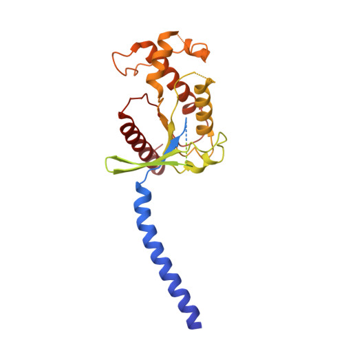
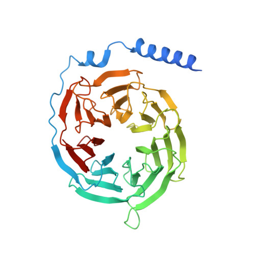
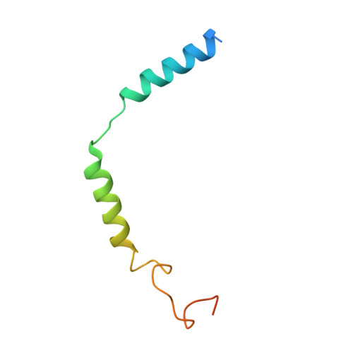
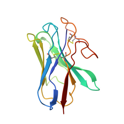
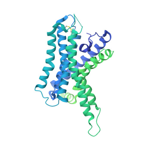
    Adhesion G protein-coupled receptors (aGPCRs) constitute an evolutionarily ancient family of receptors that often undergo autoproteolysis to produce α and β subunits 1-3 . A tethered agonism mediated by the 'Stachel sequence' of the β subunit has been proposed to have central roles in aGPCR activation 4-6 . Here we present three cryo-electron microscopy structures of aGPCRs coupled to the G s heterotrimer. Two of these aGPCRs are activated by tethered Stachel sequences-the ADGRG2-β-G s complex and the ADGRG4-β-G s complex (in which β indicates the β subunit of the aGPCR)-and the other is the full-length ADGRG2 in complex with the exogenous ADGRG2 Stachel-sequence-derived peptide agonist IP15 (ADGRG2(FL)-IP15-G s ). The Stachel sequences of both ADGRG2-β and ADGRG4-β assume a U shape and insert deeply into the seven-transmembrane bundles. Constituting the FXφφφXφ motif (in which φ represents a hydrophobic residue), five residues of ADGRG2-β or ADGRG4-β extend like fingers to mediate binding to the seven-transmembrane domain and activation of the receptor. The structure of the ADGRG2(FL)-IP15-G s complex reveals the structural basis for the improved binding affinity of IP15 compared with VPM-p15 and indicates that rational design of peptidic agonists could be achieved by exploiting aGPCR-β structures. By converting the 'finger residues' to acidic residues, we develop a method to generate peptidic antagonists towards several aGPCRs. Collectively, our study provides structural and biochemical insights into the tethered activation mechanism of aGPCRs.


  • Organizational Affiliation

    Department of Clinical Laboratory, The Second Hospital, and Advanced Medical Research Institute, Cheeloo College of Medicine, Shandong University, Jinan, China.


Macromolecules
Find similar proteins by:  (by identity cutoff)  |  3D Structure
Entity ID: 1
MoleculeChains Sequence LengthOrganismDetailsImage
Guanine nucleotide-binding protein G(s) subunit alpha isoforms short394Homo sapiensMutation(s): 8 
Gene Names: GNASGNAS1GSP
Membrane Entity: Yes 
UniProt & NIH Common Fund Data Resources
Find proteins for P63092 (Homo sapiens)
Explore P63092 
Go to UniProtKB:  P63092
PHAROS:  P63092
GTEx:  ENSG00000087460 
Entity Groups  
Sequence Clusters30% Identity50% Identity70% Identity90% Identity95% Identity100% Identity
UniProt GroupP63092
Sequence Annotations
Expand
  • Reference Sequence
Find similar proteins by:  (by identity cutoff)  |  3D Structure
Entity ID: 2
MoleculeChains Sequence LengthOrganismDetailsImage
Guanine nucleotide-binding protein G(I)/G(S)/G(T) subunit beta-1358Homo sapiensMutation(s): 0 
Gene Names: GNB1
UniProt & NIH Common Fund Data Resources
Find proteins for P62873 (Homo sapiens)
Explore P62873 
Go to UniProtKB:  P62873
PHAROS:  P62873
GTEx:  ENSG00000078369 
Entity Groups  
Sequence Clusters30% Identity50% Identity70% Identity90% Identity95% Identity100% Identity
UniProt GroupP62873
Sequence Annotations
Expand
  • Reference Sequence
Find similar proteins by:  (by identity cutoff)  |  3D Structure
Entity ID: 3
MoleculeChains Sequence LengthOrganismDetailsImage
Guanine nucleotide-binding protein G(I)/G(S)/G(O) subunit gamma-2C [auth G]71Homo sapiensMutation(s): 0 
Gene Names: GNG2
Membrane Entity: Yes 
UniProt & NIH Common Fund Data Resources
Find proteins for P59768 (Homo sapiens)
Explore P59768 
Go to UniProtKB:  P59768
PHAROS:  P59768
GTEx:  ENSG00000186469 
Entity Groups  
Sequence Clusters30% Identity50% Identity70% Identity90% Identity95% Identity100% Identity
UniProt GroupP59768
Sequence Annotations
Expand
  • Reference Sequence
Find similar proteins by:  (by identity cutoff)  |  3D Structure
Entity ID: 4
MoleculeChains Sequence LengthOrganismDetailsImage
Nanobody-35D [auth N]128Lama glamaMutation(s): 0 
Entity Groups  
Sequence Clusters30% Identity50% Identity70% Identity90% Identity95% Identity100% Identity
Sequence Annotations
Expand
  • Reference Sequence
Find similar proteins by:  (by identity cutoff)  |  3D Structure
Entity ID: 5
MoleculeChains Sequence LengthOrganismDetailsImage
Adhesion G-protein coupled receptor G2,mCherryE [auth R]683Mus musculusMutation(s): 0 
Gene Names: Adgrg2Gpr64Me6
Membrane Entity: Yes 
UniProt
Find proteins for Q8CJ12 (Mus musculus)
Explore Q8CJ12 
Go to UniProtKB:  Q8CJ12
Entity Groups  
Sequence Clusters30% Identity50% Identity70% Identity90% Identity95% Identity100% Identity
UniProt GroupQ8CJ12
Sequence Annotations
Expand
  • Reference Sequence
Experimental Data & Validation

Experimental Data

  • Method: ELECTRON MICROSCOPY
  • Resolution: 2.90 Å
  • Aggregation State: PARTICLE 
  • Reconstruction Method: SINGLE PARTICLE 

Structure Validation

View Full Validation Report


Entry History & Funding Information

Deposition Data


Funding OrganizationLocationGrant Number
National Natural Science Foundation of China (NSFC)China31971195
National Natural Science Foundation of China (NSFC)China11922410

Revision History  (Full details and data files)

  • Version 1.0: 2022-04-27
    Type: Initial release
  • Version 1.1: 2022-05-11
    Changes: Database references