Structural and functional characterization of a mycobacterial methylenetetrahydrofolate reductase utilizing NADH as the exclusive cofactor.
Li, J., Yang, M., Li, W., Lu, C., Feng, D., Shang, Z., Wang, C., Lin, W.(2023) Biochem J 480: 1129-1146
- PubMed: 37435857 Search on PubMed
- DOI: https://doi.org/10.1042/BCJ20230138
- Primary Citation Related Structures:
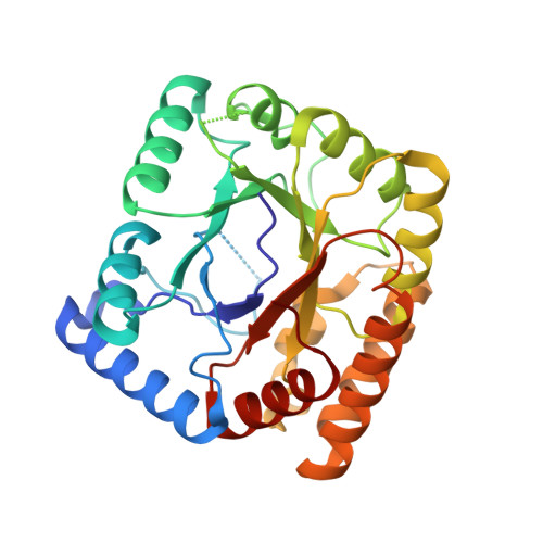
7WMW, 7WMX, 7WMY, 7WMZ - PubMed Abstract:
5,10-Methylenetetraydrofolate reductase (MTHFR) is a key enzyme in folate metabolism. MSMEG_6649, a non-canonical MTHFR from Mycobacterium smegmatis, was previously reported as a monomeric protein lacking the flavin coenzyme. However, the structural basis for its unique flavin-independent catalytic mechanism remains poorly understood. Here, we determined the crystal structures of apo MTHFR MSMEG_6649 and its complex with NADH from M. smegmatis. Structural analysis revealed that the groove formed by the loops 4 and 5 of non-canonical MSMEG_6649 interacting with FAD was significantly larger than that of canonical MTHFR. Meanwhile, the NADH-binding site in MSMEG_6649 is highly similar to the FAD binding site in canonical MTHFR, suggesting that NADH plays the same role (immediate hydride donor for methylenetetraydrofolate) as FAD in the catalytic reaction. Using biochemical analysis, molecular modeling, and site-directed mutagenesis, the critical residues participating in the binding of NADH and the substrate 5,10-methylenetetrahydrofolate as well as the product 5-methyltetrahydrofolate were identified and validated. Taken together, this work not only provides a good starting point for understanding the potential catalytic mechanism for MSMEG_6649, but also identifies an exploitable target for the development of anti-mycobacterial drugs.
- Department of Pathogen Biology, School of Medicine and Holistic Integrative Medicine, Nanjing University of Chinese Medicine, Nanjing, China.
Organizational Affiliation: