7WKZ | pdb_00007wkz

Crystal structure of the HSA complex with mycophenolate and aripiprazole


Experimental Data Snapshot

  • Method: X-RAY DIFFRACTION
  • Resolution: 2.99 Å
  • R-Value Free: 
    0.246 (Depositor), 0.239 (DCC) 
  • R-Value Work: 
    0.196 (Depositor), 0.197 (DCC) 

Starting Model: experimental
View more details

wwPDB Validation 3D Report Full Report

Validation slider image for 7WKZ

Ligand Structure Quality Assessment 


This is version 1.3 of the entry. See complete history

Literature

Structural Basis of the Change in the Interaction Between Mycophenolic Acid and Subdomain IIA of Human Serum Albumin During Renal Failure.

Yamasaki, K.Teshima, H.Yukizawa, R.Kuyama, K.Tsukigawa, K.Nishi, K.Otagiri, M.Kawai, A.

(2023) J Med Chem 66: 951-961

  • DOI: https://doi.org/10.1021/acs.jmedchem.2c01790
  • Primary Citation Related Structures: 
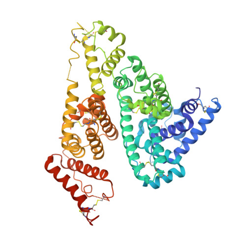
    7WKZ

  • PubMed Abstract: 

    Mycophenolic acid (MP) is an active metabolite of mycophenolate mofetil, a widely used immunosuppressive drug. MP normally exhibits high plasma protein binding (97-99%), but its binding rate is decreased in patients with renal insufficiency. This decreased protein binding is thought to be associated with leukopenia, a side effect of MP. In this study, we characterized the change in protein binding of MP in renal failure patients. Our findings indicate that MP binds strongly to subdomain IIA of human serum albumin. X-ray crystallographic data indicated that the isobenzofuran group of MP forms a stacking interaction with Trp214, and the carboxyl group of MP is located at a position that allows the formation of hydrogen bonds with Tyr150, His242, or Arg257. Due to the specific binding of MP to subdomain IIA, MP is thought to be displaced by uremic toxin (3-carboxy-4-methyl-5-propyl-2-furan-propionic acid) and fatty acids (oleate or myristate) that can bind to subdomain IIA, resulting in the decreased plasma protein binding of MP in renal failure.


  • Organizational Affiliation
    • Faculty of Pharmaceutical Sciences, Sojo University, Kumamoto860-0082, Japan.

Macromolecule Content 

  • Total Structure Weight: 134.68 kDa 
  • Atom Count: 8,443 
  • Modeled Residue Count: 1,105 
  • Deposited Residue Count: 1,170 
  • Unique protein chains: 1

Macromolecules

Find similar proteins by:|  3D Structure
Entity ID: 1
MoleculeChains  Sequence LengthOrganismDetailsImage
Serum albumin
A, B
585Homo sapiensMutation(s): 0 
Gene Names: ALBGIG20GIG42PRO0903PRO1708PRO2044PRO2619PRO2675UNQ696/PRO1341
UniProt & NIH Common Fund Data Resources
Find proteins for P02768 (Homo sapiens)
Explore P02768 
Go to UniProtKB:  P02768
PHAROS:  P02768
GTEx:  ENSG00000163631 
Entity Groups
Sequence Clusters30% Identity50% Identity70% Identity90% Identity95% Identity100% Identity
UniProt GroupP02768
Sequence Annotations
Expand
Reference Sequence

Experimental Data & Validation

Experimental Data

  • Method: X-RAY DIFFRACTION
  • Resolution: 2.99 Å
  • R-Value Free:  0.246 (Depositor), 0.239 (DCC) 
  • R-Value Work:  0.196 (Depositor), 0.197 (DCC) 
Space Group: P 1 21 1
Unit Cell:
Length ( Å )Angle ( ˚ )
a = 58.998α = 90
b = 183.182β = 106.514
c = 59.175γ = 90
Software Package:
Software NamePurpose
REFMACrefinement
XDSdata reduction
XDSdata scaling
MOLREPphasing

Structure Validation

View Full Validation Report


Ligand Structure Quality Assessment 


Entry History 

& Funding Information

Deposition Data


Funding OrganizationLocationGrant Number
Not funded--

Revision History  (Full details and data files)

  • Version 1.0: 2022-12-28
    Type: Initial release
  • Version 1.1: 2023-01-25
    Changes: Database references
  • Version 1.2: 2023-11-29
    Changes: Data collection, Refinement description
  • Version 1.3: 2024-11-06
    Changes: Structure summary