


Crystal structure of the adenylation domain from an epsilon-poly-l-lysine synthetase provides molecular mechanism for substrate specificity
Okamoto, T., Yamanaka, K., Hamano, Y., Nagano, S., Hino, T.(2022) Biochem Biophys Res Commun 596: 43-48
Experimental Data Snapshot
Starting Model: experimental
View more details
(2022) Biochem Biophys Res Commun 596: 43-48
Entity ID: 1 | |||||
|---|---|---|---|---|---|
| Molecule | Chains | Sequence Length | Organism | Details | Image |
| Epsilon-poly-L-lysine synthase | 509 | Streptomyces noursei | Mutation(s): 0 Gene Names: pls | 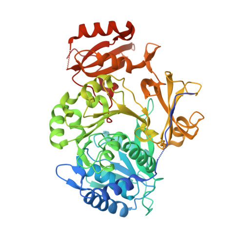 | |
UniProt | |||||
Entity Groups | |||||
| Sequence Clusters | 30% Identity50% Identity70% Identity90% Identity95% Identity100% Identity | ||||
| UniProt Group | B5BR95 | ||||
Sequence AnnotationsExpand | |||||
Reference Sequence | |||||
| Ligands 3 Unique | |||||
|---|---|---|---|---|---|
| ID | Chains | Name / Formula / InChI Key | 2D Diagram | 3D Interactions | |
| LAD (Subject of Investigation/LOI) Download:Ideal Coordinates CCD File | K [auth A] | ADENOSINE-5'-[LYSYL-PHOSPHATE] C16 H26 N7 O8 P RZWIOOBQBMRZTQ-OPYVMVOTSA-N |  | ||
| SO4 Download:Ideal Coordinates CCD File | B [auth A] C [auth A] D [auth A] E [auth A] F [auth A] | SULFATE ION O4 S QAOWNCQODCNURD-UHFFFAOYSA-L |  | ||
| GOL Download:Ideal Coordinates CCD File | H [auth A], I [auth A], J [auth A] | GLYCEROL C3 H8 O3 PEDCQBHIVMGVHV-UHFFFAOYSA-N |  | ||
| Length ( Å ) | Angle ( ˚ ) |
|---|---|
| a = 152.511 | α = 90 |
| b = 152.511 | β = 90 |
| c = 48.202 | γ = 120 |
| Software Name | Purpose |
|---|---|
| PHENIX | refinement |
| XDS | data reduction |
| Aimless | data scaling |
| PHASER | phasing |
| Coot | model building |
| Funding Organization | Location | Grant Number |
|---|---|---|
| Japan Society for the Promotion of Science (JSPS) | Japan | 20H02918 |
| Japan Society for the Promotion of Science (JSPS) | Japan | 16H06445 |
| Japan Society for the Promotion of Science (JSPS) | Japan | 19K06551 |


