In situ crystal data-collection and ligand-screening system at SPring-8.
Okumura, H., Sakai, N., Murakami, H., Mizuno, N., Nakamura, Y., Ueno, G., Masunaga, T., Kawamura, T., Baba, S., Hasegawa, K., Yamamoto, M., Kumasaka, T.(2022) Acta Crystallogr F Struct Biol Commun 78: 241-251
- PubMed: 35647681 Search on PubMedSearch on PubMed Central
- DOI: https://doi.org/10.1107/S2053230X22005283
- Primary Citation Related Structures:
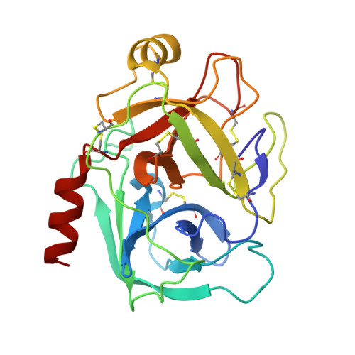
7WA0, 7WA2, 7WB6, 7WB7, 7WB8, 7WB9, 7WBA - PubMed Abstract:
In situ diffraction data collection using crystallization plates has been utilized for macromolecules to evaluate crystal quality without requiring additional sample treatment such as cryocooling. Although it is difficult to collect complete data sets using this technique due to the mechanical limitation of crystal rotation, recent advances in methods for data collection from multiple crystals have overcome this issue. At SPring-8, an in situ diffraction measurement system was constructed consisting of a goniometer for a plate, an articulated robot and plate storage. Using this system, complete data sets were obtained utilizing the small-wedge measurement method. Combining this system with an acoustic liquid handler to prepare protein-ligand complex crystals by applying fragment compounds to trypsin crystals for in situ soaking, binding was confirmed for seven out of eight compounds. These results show that the system functioned properly to collect complete data for structural analysis and to expand the capability for ligand screening in combination with a liquid dispenser.
- Structural Biology Division, Japan Synchrotron Radiation Research Institute, 1-1-1 Kouto, Sayo-cho, Sayo-gun, Hyogo 679-5198, Japan.
Organizational Affiliation: