7W86 | pdb_00007w86

Crystal structure of the DYW domain of DYW1


Experimental Data Snapshot

  • Method: X-RAY DIFFRACTION
  • Resolution: 1.80 Å
  • R-Value Free: 
    0.234 (Depositor), 0.234 (DCC) 
  • R-Value Work: 
    0.177 (Depositor), 0.192 (DCC) 
  • R-Value Observed: 
    0.180 (Depositor) 

wwPDB Validation 3D Report Full Report

Validation slider image for 7W86

This is version 1.2 of the entry. See complete history

Literature

Structural insight into the activation of an Arabidopsis organellar C-to-U RNA editing enzyme by active site complementation.

Toma-Fukai, S.Sawada, Y.Maeda, A.Shimizu, H.Shikanai, T.Takenaka, M.Shimizu, T.

(2023) Plant Cell 35: 1888-1900

  • DOI: https://doi.org/10.1093/plcell/koac318
  • Primary Citation Related Structures: 
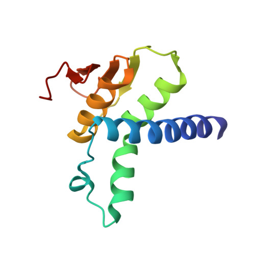
    7W86

  • PubMed Abstract: 

    RNA-binding pentatricopeptide repeat (PPR) proteins catalyze hundreds of cytidine to uridine RNA editing events in plant organelles; these editing events are essential for proper gene expression. More than half of the PPR-type RNA editing factors, however, lack the DYW cytidine deaminase domain. Genetic analyses have suggested that their cytidine deaminase activity arises by association with a family of DYW1-like proteins that contain an N-terminally truncated DYW domain, but their molecular mechanism has been unclear. Here, we report the crystal structure of the Arabidopsis thaliana DYW1 deaminase domain at 1.8 Å resolution. DYW1 has a cytidine deaminase fold lacking the PG box. The internal insertion within the deaminase fold shows an α-helical fold instead of the β-finger reported for the gating domain of the A. thaliana ORGANELLE TRANSCRIPT PROCESSING 86. The substrate-binding pocket is incompletely formed and appears to be complemented in the complex by the E2 domain and the PG box of the interacting PPR protein. In vivo RNA editing assays corroborate the activation model for DYW1 deaminase. Our study demonstrates the common activation mechanism of the DYW1-like proteins by molecular complementation of the DYW domain and reconstitution of the substrate-binding pocket.


  • Organizational Affiliation
    • Graduate School of Pharmaceutical Sciences, The University of Tokyo, 7-3-1 Hongo, Bunkyo-ku, Tokyo 113-0033, Japan.

Macromolecule Content 

  • Total Structure Weight: 14.97 kDa 
  • Atom Count: 1,066 
  • Modeled Residue Count: 117 
  • Deposited Residue Count: 117 
  • Unique protein chains: 1

Macromolecules

Find similar proteins by:|  3D Structure
Entity ID: 1
MoleculeChains  Sequence LengthOrganismDetailsImage
Pentatricopeptide repeat-containing protein DWY1, chloroplastic117Arabidopsis thalianaMutation(s): 0 
Gene Names: DYW1PCMP-H50At1g47580F16N3.14
UniProt
Find proteins for P0C7R1 (Arabidopsis thaliana)
Explore P0C7R1 
Go to UniProtKB:  P0C7R1
Entity Groups
Sequence Clusters30% Identity50% Identity70% Identity90% Identity95% Identity100% Identity
UniProt GroupP0C7R1
Sequence Annotations
Expand
Reference Sequence

Small Molecules

Ligands 5 Unique
IDChains Name / Formula / InChI Key2D Diagram3D Interactions
SO4

Query on SO4


Download:Ideal Coordinates CCD File
E [auth A]SULFATE ION
O4 S
QAOWNCQODCNURD-UHFFFAOYSA-L
GOL

Query on GOL


Download:Ideal Coordinates CCD File
F [auth A],
K [auth A],
Q [auth A],
T [auth A]
GLYCEROL
C3 H8 O3
PEDCQBHIVMGVHV-UHFFFAOYSA-N
ZN
(Subject of Investigation/LOI)

Query on ZN


Download:Ideal Coordinates CCD File
B [auth A],
C [auth A],
D [auth A]
ZINC ION
Zn
PTFCDOFLOPIGGS-UHFFFAOYSA-N
EDO

Query on EDO


Download:Ideal Coordinates CCD File
G [auth A]
I [auth A]
L [auth A]
M [auth A]
N [auth A]
G [auth A],
I [auth A],
L [auth A],
M [auth A],
N [auth A],
P [auth A],
R [auth A],
S [auth A],
U [auth A],
V [auth A]
1,2-ETHANEDIOL
C2 H6 O2
LYCAIKOWRPUZTN-UHFFFAOYSA-N
ACT

Query on ACT


Download:Ideal Coordinates CCD File
H [auth A],
J [auth A],
O [auth A]
ACETATE ION
C2 H3 O2
QTBSBXVTEAMEQO-UHFFFAOYSA-M

Experimental Data & Validation

Experimental Data

  • Method: X-RAY DIFFRACTION
  • Resolution: 1.80 Å
  • R-Value Free:  0.234 (Depositor), 0.234 (DCC) 
  • R-Value Work:  0.177 (Depositor), 0.192 (DCC) 
  • R-Value Observed: 0.180 (Depositor) 
Space Group: P 1 21 1
Unit Cell:
Length ( Å )Angle ( ˚ )
a = 32.945α = 90
b = 45.165β = 98.04
c = 37.023γ = 90
Software Package:
Software NamePurpose
REFMACrefinement
XDSdata reduction
Aimlessdata scaling
MOLREPphasing

Structure Validation

View Full Validation Report


Entry History 

& Funding Information

Deposition Data


Funding OrganizationLocationGrant Number
Ministry of Education, Culture, Sports, Science and Technology (Japan)Japan--
Japan Agency for Medical Research and Development (AMED)Japan--
Japan Science and TechnologyJapan--

Revision History  (Full details and data files)

  • Version 1.0: 2022-12-14
    Type: Initial release
  • Version 1.1: 2023-06-28
    Changes: Database references
  • Version 1.2: 2024-05-29
    Changes: Data collection