7W7W

E2 Pi of SERCA2b


Experimental Data Snapshot

  • Method: ELECTRON MICROSCOPY
  • Resolution: 3.20 Å
  • Aggregation State: PARTICLE 
  • Reconstruction Method: SINGLE PARTICLE 

wwPDB Validation   3D Report Full Report


This is version 1.1 of the entry. See complete history


Literature

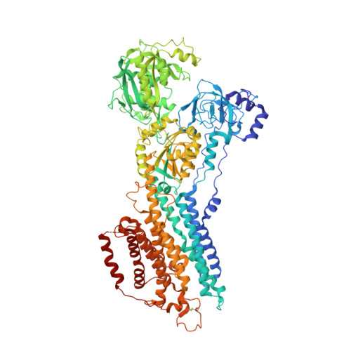
Multiple sub-state structures of SERCA2b reveal conformational overlap at transition steps during the catalytic cycle.

Zhang, Y.Kobayashi, C.Cai, X.Watanabe, S.Tsutsumi, A.Kikkawa, M.Sugita, Y.Inaba, K.

(2022) Cell Rep 41: 111760-111760

  • DOI: https://doi.org/10.1016/j.celrep.2022.111760
  • Primary Citation of Related Structures:  
    7W7T, 7W7U, 7W7V, 7W7W

  • PubMed Abstract: 

    Sarco/endoplasmic reticulum Ca 2+ ATPase (SERCA) pumps Ca 2+ into the endoplasmic reticulum (ER). Herein, we present cryo-electron microscopy (EM) structures of three intermediates of SERCA2b: Ca 2+ -bound phosphorylated (E1P·2Ca 2+ ) and Ca 2+ -unbound dephosphorylated (E2·Pi) intermediates and another between the E2P and E2·Pi states. Our cryo-EM analysis demonstrates that the E1P·2Ca 2+ state exists in low abundance and preferentially transitions to an E2P-like structure by releasing Ca 2+ and that the Ca 2+ release gate subsequently undergoes stepwise closure during the dephosphorylation processes. Importantly, each intermediate adopts multiple sub-state structures including those like the next one in the catalytic series, indicating conformational overlap at transition steps, as further substantiated by atomistic molecular dynamic simulations of SERCA2b in a lipid bilayer. The present findings provide insight into how enzymes accelerate catalytic cycles.


  • Organizational Affiliation

    Institute of Multidisciplinary Research for Advanced Materials, Tohoku University, Katahira 2-1-1, Aoba-ku, Sendai 980-8577, Japan.


Macromolecules
Find similar proteins by:  (by identity cutoff)  |  3D Structure
Entity ID: 1
MoleculeChains Sequence LengthOrganismDetailsImage
Sarcoplasmic/endoplasmic reticulum calcium ATPase 21,042Homo sapiensMutation(s): 0 
Gene Names: ATP2A2ATP2B
EC: 7.2.2.10
Membrane Entity: Yes 
UniProt & NIH Common Fund Data Resources
Find proteins for P16615 (Homo sapiens)
Explore P16615 
Go to UniProtKB:  P16615
PHAROS:  P16615
GTEx:  ENSG00000174437 
Entity Groups  
Sequence Clusters30% Identity50% Identity70% Identity90% Identity95% Identity100% Identity
UniProt GroupP16615
Sequence Annotations
Expand
  • Reference Sequence
Experimental Data & Validation

Experimental Data

  • Method: ELECTRON MICROSCOPY
  • Resolution: 3.20 Å
  • Aggregation State: PARTICLE 
  • Reconstruction Method: SINGLE PARTICLE 

Structure Validation

View Full Validation Report


Entry History & Funding Information

Deposition Data


Funding OrganizationLocationGrant Number
Ministry of Education, Culture, Sports, Science and Technology (Japan)Japan18H03978
Ministry of Education, Culture, Sports, Science and Technology (Japan)Japan21H04758
Ministry of Education, Culture, Sports, Science and Technology (Japan)Japan21H05247
Ministry of Education, Culture, Sports, Science and Technology (Japan)Japan21K15036

Revision History  (Full details and data files)

  • Version 1.0: 2022-12-14
    Type: Initial release
  • Version 1.1: 2023-01-11
    Changes: Database references, Structure summary