7W7R

High resolution structure of a fish aquaporin reveals a novel extracellular fold.


Experimental Data Snapshot

  • Method: X-RAY DIFFRACTION
  • Resolution: 3.46 Å
  • R-Value Free: 0.299 
  • R-Value Work: 0.271 
  • R-Value Observed: 0.272 

wwPDB Validation   3D Report Full Report


This is version 1.2 of the entry. See complete history


Literature

High-resolution structure of a fish aquaporin reveals a novel extracellular fold.

Zeng, J.Schmitz, F.Isaksson, S.Glas, J.Arbab, O.Andersson, M.Sundell, K.Eriksson, L.A.Swaminathan, K.Tornroth-Horsefield, S.Hedfalk, K.

(2022) Life Sci Alliance 5

  • DOI: https://doi.org/10.26508/lsa.202201491
  • Primary Citation of Related Structures:  
    7W7R, 7W7S

  • PubMed Abstract: 

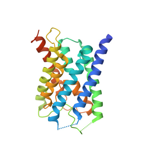
    Aquaporins are protein channels embedded in the lipid bilayer in cells from all organisms on earth that are crucial for water homeostasis. In fish, aquaporins are believed to be important for osmoregulation; however, the molecular mechanism behind this is poorly understood. Here, we present the first structural and functional characterization of a fish aquaporin; cpAQP1aa from the fresh water fish climbing perch (Anabas testudineus), a species that is of high osmoregulatory interest because of its ability to spend time in seawater and on land. These studies show that cpAQP1aa is a water-specific aquaporin with a unique fold on the extracellular side that results in a constriction region. Functional analysis combined with molecular dynamic simulations suggests that phosphorylation at two sites causes structural perturbations in this region that may have implications for channel gating from the extracellular side.


  • Organizational Affiliation

    Department of Biological Sciences, National University of Singapore, Queenstown, Singapore.


Macromolecules
Find similar proteins by:  (by identity cutoff)  |  3D Structure
Entity ID: 1
MoleculeChains Sequence LengthOrganismDetailsImage
Aquaporin 1
A, B, C, D
244Anabas testudineusMutation(s): 0 
Gene Names: aqp1aaaqp1
Membrane Entity: Yes 
UniProt
Find proteins for M1K561 (Anabas testudineus)
Explore M1K561 
Go to UniProtKB:  M1K561
Entity Groups  
Sequence Clusters30% Identity50% Identity70% Identity90% Identity95% Identity100% Identity
UniProt GroupM1K561
Sequence Annotations
Expand
  • Reference Sequence
Experimental Data & Validation

Experimental Data

  • Method: X-RAY DIFFRACTION
  • Resolution: 3.46 Å
  • R-Value Free: 0.299 
  • R-Value Work: 0.271 
  • R-Value Observed: 0.272 
  • Space Group: C 2 2 21
Unit Cell:
Length ( Å )Angle ( ˚ )
a = 113.67α = 90
b = 178.1β = 90
c = 177.9γ = 90
Software Package:
Software NamePurpose
SCALAdata scaling
PHENIXrefinement
PDB_EXTRACTdata extraction
iMOSFLMdata reduction
PHASERphasing

Structure Validation

View Full Validation Report


Entry History & Funding Information

Deposition Data


Funding OrganizationLocationGrant Number
Swedish Research CouncilSwedenK2013-66X-20431-07-05
Swedish Research CouncilSweden2009-00360
Swedish Research CouncilSweden2013-05945
Ministry of Education (MoE, Singapore)SingaporeR154-000-632-112
National Research Foundation (NRF, Singapore)SingaporeR706-000-041-279
Swedish Research Council FORMASSweden2012-771

Revision History  (Full details and data files)

  • Version 1.0: 2022-10-12
    Type: Initial release
  • Version 1.1: 2023-04-26
    Changes: Database references
  • Version 1.2: 2023-11-29
    Changes: Data collection, Refinement description