7VZ3 | pdb_00007vz3

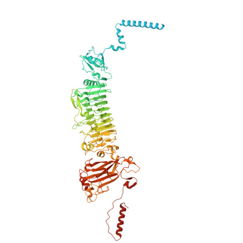
Cryo-EM structure of Depo32, a Klebsiella phage depolymerase targets the K2 serotype K. pneumoniae


Experimental Data Snapshot

  • Method: ELECTRON MICROSCOPY
  • Resolution: 2.46 Å
  • Aggregation State: PARTICLE 
  • Reconstruction Method: SINGLE PARTICLE 

wwPDB Validation 3D Report Full Report

Validation slider image for 7VZ3

This is version 1.1 of the entry. See complete history. 

Literature

Structural biology and functional features of phage-derived depolymerase Depo32 on Klebsiella pneumoniae with K2 serotype capsular polysaccharides.

Cai, R., Ren, Z., Zhao, R., Lu, Y., Wang, X., Guo, Z., Song, J., Xiang, W., Du, R., Zhang, X., Han, W., Ru, H., Gu, J.

(2023) Microbiol Spectr 11: e0530422-e0530422

  • DOI: https://doi.org/10.1128/spectrum.05304-22
  • Primary Citation Related Structures: 
    7VYV, 7VZ3

  • PubMed Abstract: 

    Depolymerases specific to more than 20 serotypes of Klebsiella spp. have been identified, but most studies only evaluated the single-dose treatment of depolymerases with relatively simple clinical evaluation indices and did not reveal the anti-infection mechanism of these depolymerases in depth. On the basis of determining the biological characteristics, the structure of Depo32 was analyzed by cryo-electron microscopy, and the potential active center was further identified. In addition, the effects of Depo32 on macrophage phagocytosis, signaling pathway activation, and serum killing were revealed, and the efficacy of the depolymerase (single treatment, multiple treatments, or in combination with gentamicin) against acute pneumonia caused by Klebsiella pneumoniae was evaluated. Moreover, the roles of the active sites of Depo32 were also elucidated in the in vitro and in vivo studies. Therefore, through structural biology, cell biology, and in vivo experiments, this study demonstrated the mechanism by which Depo32 targets K2 serotype K . pneumoniae infection.


  • Organizational Affiliation: 
    • College of Animal Science and Technology, Jilin Agricultural University , Changchun, Jilin, China.

Macromolecule Content 

  • Total Structure Weight: 302.84 kDa 
  • Atom Count: 17,415 
  • Modeled Residue Count: 2,256 
  • Deposited Residue Count: 2,784 
  • Unique protein chains: 1

Macromolecules

Find similar proteins by:|  3D Structure
Entity ID: 1
MoleculeChains  Sequence LengthOrganismDetailsImage
Depolymerase
A, B, C
928Klebsiella phage GH-K3Mutation(s): 0 
Gene Names: GHK3_32
UniProt
Find proteins for A0A3S7W7I3 (Klebsiella phage GH-K3)
Explore A0A3S7W7I3 
Go to UniProtKB:  A0A3S7W7I3
Entity Groups
Sequence Clusters30% Identity50% Identity70% Identity90% Identity95% Identity100% Identity
UniProt GroupA0A3S7W7I3
Sequence Annotations
Expand
Reference Sequence

Experimental Data & Validation

Experimental Data

  • Method: ELECTRON MICROSCOPY
  • Resolution: 2.46 Å
  • Aggregation State: PARTICLE 
  • Reconstruction Method: SINGLE PARTICLE 
EM Software:
TaskSoftware PackageVersion
RECONSTRUCTIONRELION
MODEL REFINEMENTPHENIX

Structure Validation

View Full Validation Report


Entry History 

& Funding Information

Deposition Data


Funding OrganizationLocationGrant Number
National Natural Science Foundation of China (NSFC)China32072824
National Natural Science Foundation of China (NSFC)China31872505
National Natural Science Foundation of China (NSFC)ChinaU19A2038

Revision History  (Full details and data files)

  • Version 1.0: 2023-08-30
    Type: Initial release
  • Version 1.1: 2024-09-11
    Changes: Data collection, Database references