Snapshots of urea-induced early structural changes and unfolding of an ankyrin repeat protein at atomic resolution.
Medur Gurushankar, M.S., Dalvi, S., Venkatraman, P.(2022) Protein Sci 31: e4515-e4515
- PubMed: 36382986 Search on PubMedSearch on PubMed Central
- DOI: https://doi.org/10.1002/pro.4515
- Primary Citation Related Structures:
7VXV, 7VXW, 7VY4, 7VY7 - PubMed Abstract:
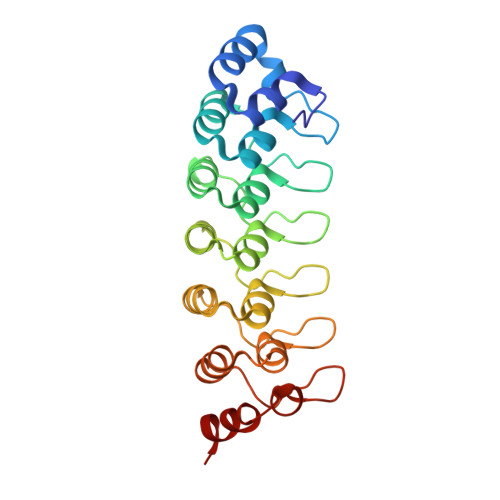
Protein folding and unfolding is a complex process, underscored by the many proteotoxic diseases associated with misfolded proteins. Mapping pathways from a native structure to an unfolded protein or vice versa, identifying the intermediates, and defining the role of sequence and structure en route remain outstanding problems in the field. It is even more challenging to capture the events at atomistic resolution. X-ray diffraction has so far been used to understand how urea interacts with and unfolds two stable globular proteins. Here, we present the case study on PSMD10 Gankyrin , a prototype for a moderately stable, non-globular repeat protein, long and rigid, with its termini located at either end. We define structural changes in the time dimension using low urea concentrations to arrive at the following conclusions. (a) Unfolding is rapidly initiated at the C-terminus, slowly at the N-terminus, and proceeds inwards from both ends. (b) C-terminus undergoes an α to 3 10 helix transition, representing the structure of a potential early unfolding intermediate before disorder sets in. (c) Distinct and progressive changes in the electrostatic landscape of PSMD10 Gankyrin were observed, indicative of conformational changes in the seemingly inflexible motif involved in protein-protein interaction. We believe this unique study will open up the field for better and bolder queries and increase the choice of model proteins for a better understanding of the challenging problems of protein folding, protein interactions, protein degradation, and diseases associated with misfolding.
- Protein Interactome Laboratory for Structural and Functional Biology, Advanced Centre for Treatment, Research and Education in Cancer, Navi Mumbai, Maharashtra, India.
Organizational Affiliation: