


Chitporin from Escherichia coli complex with chitohexaose
Soysa, H.S.M., Suginta, W., Amornloetwattana, R., van den Berg, B.To be published.
Experimental Data Snapshot
Starting Model: experimental
View more details
Entity ID: 1 | |||||
|---|---|---|---|---|---|
| Molecule | Chains | Sequence Length | Organism | Details | Image |
| Chitoporin | 436 | Escherichia coli K-12 | Mutation(s): 0 Gene Names: chiP, ybfM, b0681, JW0667 Membrane Entity: Yes | 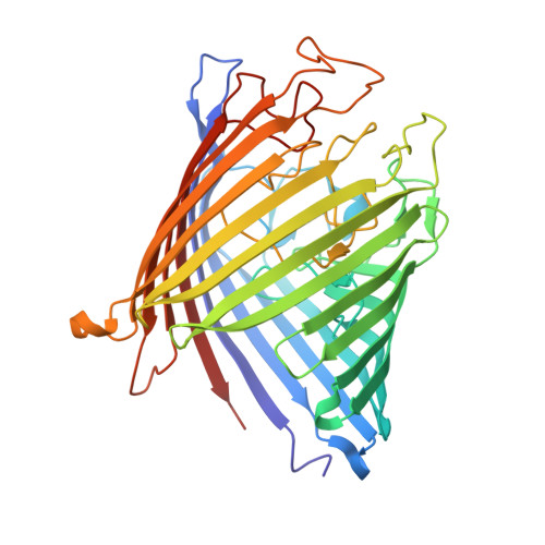 | |
UniProt | |||||
Entity Groups | |||||
| Sequence Clusters | 30% Identity50% Identity70% Identity90% Identity95% Identity100% Identity | ||||
| UniProt Group | P75733 | ||||
Sequence AnnotationsExpand | |||||
Reference Sequence | |||||
Entity ID: 2 | |||||
|---|---|---|---|---|---|
| Molecule | Chains | Length | 2D Diagram | Glycosylation | D Interactions |
| 2-acetamido-2-deoxy-beta-D-glucopyranose-(1-4)-2-acetamido-2-deoxy-beta-D-glucopyranose-(1-4)-2-acetamido-2-deoxy-beta-D-glucopyranose-(1-4)-2-acetamido-2-deoxy-beta-D-glucopyranose-(1-4)-2-acetamido-2-deoxy-beta-D-glucopyranose | C [auth U], D [auth C] | 5 |  | N/A | |
Glycosylation Resources | |||||
GlyTouCan: G07670VS GlyCosmos: G07670VS GlyGen: G07670VS | |||||
| Ligands 3 Unique | |||||
|---|---|---|---|---|---|
| ID | Chains | Name / Formula / InChI Key | 2D Diagram | 3D Interactions | |
| DMU Download:Ideal Coordinates CCD File | E [auth A] F [auth A] G [auth A] S [auth B] T [auth B] | DECYL-BETA-D-MALTOPYRANOSIDE C22 H42 O11 WOQQAWHSKSSAGF-WXFJLFHKSA-N |  | ||
| C8E Download:Ideal Coordinates CCD File | AA [auth B] BA [auth B] CA [auth B] DA [auth B] H [auth A] | (HYDROXYETHYLOXY)TRI(ETHYLOXY)OCTANE C16 H34 O5 FEOZZFHAVXYAMB-UHFFFAOYSA-N |  | ||
| MG Download:Ideal Coordinates CCD File | EA [auth B] FA [auth B] GA [auth B] HA [auth B] Q [auth A] | MAGNESIUM ION Mg JLVVSXFLKOJNIY-UHFFFAOYSA-N |  | ||
| Length ( Å ) | Angle ( ˚ ) |
|---|---|
| a = 94.737 | α = 90 |
| b = 58.551 | β = 110.708 |
| c = 133.059 | γ = 90 |
| Software Name | Purpose |
|---|---|
| PHENIX | refinement |
| xia2 | data reduction |
| Aimless | data scaling |
| PHASER | phasing |
| Funding Organization | Location | Grant Number |
|---|---|---|
| European Union (EU) | European Union | -- |


