Structural comparison between the DNA-protective ability of scallop and shrimp ferritin from iron-induced oxidative damage.
Zhang, C., Liu, Y., Zhang, T., Lv, C., Zang, J., Zhao, G.(2022) Food Chem 386: 132827-132827
- PubMed: 35364499 Search on PubMed
- DOI: https://doi.org/10.1016/j.foodchem.2022.132827
- Primary Citation Related Structures:
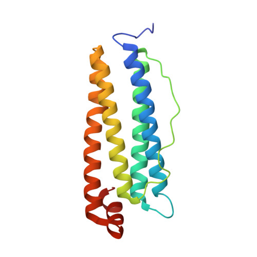
7VT2 - PubMed Abstract:
The structure and function of ferritin from seafood has been largely unexplored. In this study, homopolymeric scallop ferritin (ApF) was prepared for the first time, the apo form of which exhibited the stronger ability to protect DNA from iron-induced oxidative damage as compared to its analogue, homopolymeric shrimp ferritin (MjF). Their difference in DNA-protective activity was derived from less hydroxyl radicals produced during iron oxidation in the presence of scallop ferritin than shrimp ferritin. The kinetic results showed that apo-ApF catalyzed the faster ferrous ions oxidation by oxygen into nontoxic ferric ions as compared to apo-MjF. Newly reported crystal structure of ApF revealed that its stronger ferroxidase activity stemmed from different structures in the triple axis channel and ferroxidase site as compared to MjF. All these new findings advance our understanding of the relationship between the structure and function of food-related protein.
- College of Food Science & Nutritional Engineering, China Agricultural University, Key Laboratory of Functional Dairy, Ministry of Education, Beijing 100083, China.
Organizational Affiliation: