 Download Mendeley
Cryo-EM structures of infectious bursal disease viruses with different virulences provide insights into their assembly and invasion
Bao, K., Qi, X., Lia, Y., Gong, M., Wang, M., Zhu, P.(2022) Sci Bull (Beijing) 67: 646-654
Experimental Data Snapshot
wwPDB Validation 3D Report Full Report

Entity ID: 1 | |||||
|---|---|---|---|---|---|
| Molecule | Chains | Sequence Length | Organism | Details | Image |
| Structural polyprotein | 441 | Infectious bursal disease virus | Mutation(s): 0 EC: 3.4.21 | 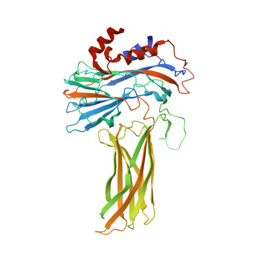 | |
UniProt | |||||
Entity Groups | |||||
| Sequence Clusters | 30% Identity50% Identity70% Identity90% Identity95% Identity100% Identity | ||||
| UniProt Group | Q9WI42 | ||||
Sequence AnnotationsExpand | |||||
Reference Sequence | |||||
| Funding Organization | Location | Grant Number |
|---|---|---|
| National Natural Science Foundation of China (NSFC) | China | 31430087 |
| National Natural Science Foundation of China (NSFC) | China | 31425007 |
