7VQQ | pdb_00007vqq

Cryo-EM structure of amyloid fibril formed by FUS low complexity domain


Experimental Data Snapshot

  • Method: ELECTRON MICROSCOPY
  • Resolution: 2.90 Å
  • Aggregation State: FILAMENT 
  • Reconstruction Method: HELICAL 

wwPDB Validation 3D Report Full Report

Validation slider image for 7VQQ

This is version 1.2 of the entry. See complete history

Literature

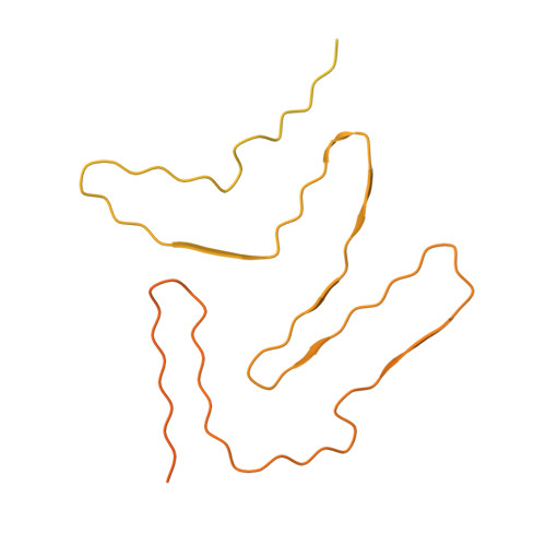
Molecular structure of an amyloid fibril formed by FUS low-complexity domain.

Sun, Y.Zhang, S.Hu, J.Tao, Y.Xia, W.Gu, J.Li, Y.Cao, Q.Li, D.Liu, C.

(2022) iScience 25: 103701-103701

  • DOI: https://doi.org/10.1016/j.isci.2021.103701
  • Primary Citation Related Structures: 
    7VQQ

  • PubMed Abstract: 

    FUS is a multifunctional nuclear protein which undergoes liquid-liquid phase separation in response to stress and DNA damage. Dysregulation of FUS dynamic phase separation leads to formation of pathological fibril closely associated with neurodegenerative diseases such as amyotrophic lateral sclerosis and frontotemporal dementia. In this study, we determined the cryo-EM structure of a cytotoxic fibril formed by the low-complexity (LC) domain of FUS at 2.9 Å resolution. The fibril structure exhibits a new and extensive serpentine fold consisting of three motifs incorporating together via a Tyr triad. FUS LC employs 91 residues to form an enlarged and stable fibril core via hydrophilic interaction and hydrogen bonds, which is distinct from most of previously determined fibrils commonly stabilized by hydrophobic interaction. Our work reveals the structural basis underlying formation of a cytotoxic and thermostable fibril of FUS LC and sheds light on understanding the liquid-to-solid phase transition of FUS in disease.


  • Organizational Affiliation
    • Interdisciplinary Research Center on Biology and Chemistry, Shanghai Institute of Organic Chemistry, Chinese Academy of Sciences, Shanghai 201210, China.

Macromolecule Content 

  • Total Structure Weight: 155.85 kDa 
  • Atom Count: 1,968 
  • Modeled Residue Count: 273 
  • Deposited Residue Count: 1,449 
  • Unique protein chains: 1

Macromolecules

Find similar proteins by:|  3D Structure
Entity ID: 1
MoleculeChains  Sequence LengthOrganismDetailsImage
fusion protein of mCerulean and FUS LCD
A, B, C
483Aequorea victoriaHomo sapiens
This entity is chimeric
Mutation(s): 0 
UniProt & NIH Common Fund Data Resources
Find proteins for P35637 (Homo sapiens)
Explore P35637 
Go to UniProtKB:  P35637
PHAROS:  P35637
GTEx:  ENSG00000089280 
Entity Groups
Sequence Clusters30% Identity50% Identity70% Identity90% Identity95% Identity100% Identity
UniProt GroupP35637
Sequence Annotations
Expand
Reference Sequence

Experimental Data & Validation

Experimental Data

  • Method: ELECTRON MICROSCOPY
  • Resolution: 2.90 Å
  • Aggregation State: FILAMENT 
  • Reconstruction Method: HELICAL 

Structure Validation

View Full Validation Report


Entry History 

& Funding Information

Deposition Data


Funding OrganizationLocationGrant Number
Not funded--

Revision History  (Full details and data files)

  • Version 1.0: 2021-12-22
    Type: Initial release
  • Version 1.1: 2022-02-23
    Changes: Database references
  • Version 1.2: 2024-06-26
    Changes: Data collection