


Crystal structure of KPC-2 beta-lactamase complexed with hydrolyzed EXW-1
Xie, H.X.To be published.
Experimental Data Snapshot
Starting Model: experimental
View more details
Entity ID: 1 | |||||
|---|---|---|---|---|---|
| Molecule | Chains | Sequence Length | Organism | Details | Image |
| Carbapenem-hydrolyzing beta-lactamase KPC | 266 | Klebsiella pneumoniae | Mutation(s): 0 Gene Names: bla, kpc, kpc1 EC: 3.5.2.6 | 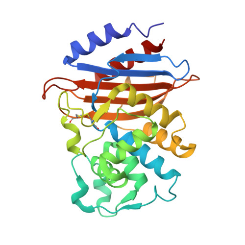 | |
UniProt | |||||
Entity Groups | |||||
| Sequence Clusters | 30% Identity50% Identity70% Identity90% Identity95% Identity100% Identity | ||||
| UniProt Group | Q9F663 | ||||
Sequence AnnotationsExpand | |||||
Reference Sequence | |||||
| Ligands 2 Unique | |||||
|---|---|---|---|---|---|
| ID | Chains | Name / Formula / InChI Key | 2D Diagram | 3D Interactions | |
| 7TC (Subject of Investigation/LOI) Download:Ideal Coordinates CCD File | T [auth C], Y [auth D] | (2R,3S)-3-methyl-4-methylidene-2-[(2S,3R)-3-oxidanyl-1-oxidanylidene-butan-2-yl]-2,3-dihydropyrrole-5-carboxylic acid C11 H15 N O4 GGYAWCWTCNLFOB-KDBVAPGDSA-N |  | ||
| SO4 Download:Ideal Coordinates CCD File | E [auth A] F [auth A] G [auth A] H [auth A] I [auth B] | SULFATE ION O4 S QAOWNCQODCNURD-UHFFFAOYSA-L |  | ||
| Length ( Å ) | Angle ( ˚ ) |
|---|---|
| a = 64.193 | α = 90 |
| b = 127.539 | β = 104.288 |
| c = 76.303 | γ = 90 |
| Software Name | Purpose |
|---|---|
| PHENIX | refinement |
| HKL-3000 | data reduction |
| HKL-3000 | data scaling |
| MOLREP | phasing |
| Funding Organization | Location | Grant Number |
|---|---|---|
| National Natural Science Foundation of China (NSFC) | China | -- |


