


Crystal structure of a hydrolase in apo form 2
Wu, P., Zhao, Y.P., Li, Z.S., Ingrid, M.C., Lara, P., Gao, J., Han, X., Li, Q., Basak, O., Liu, W.D., Wei, R.To be published.
Experimental Data Snapshot
Starting Model: experimental
View more details
wwPDB Validation 3D Report Full Report

Entity ID: 1 | |||||
|---|---|---|---|---|---|
| Molecule | Chains | Sequence Length | Organism | Details | Image |
| hydrolase | 293 | unclassified Marinobacter | Mutation(s): 0 | 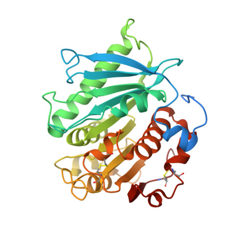 | |
UniProt | |||||
Find proteins for A0A9X9ZA41 (unclassified Marinobacter) Explore A0A9X9ZA41 Go to UniProtKB: A0A9X9ZA41 | |||||
Entity Groups | |||||
| Sequence Clusters | 30% Identity50% Identity70% Identity90% Identity95% Identity100% Identity | ||||
| UniProt Group | A0A9X9ZA41 | ||||
Sequence AnnotationsExpand | |||||
Reference Sequence | |||||
| Ligands 1 Unique | |||||
|---|---|---|---|---|---|
| ID | Chains | Name / Formula / InChI Key | 2D Diagram | 3D Interactions | |
| CA Download:Ideal Coordinates CCD File | B [auth A], C [auth A] | CALCIUM ION Ca BHPQYMZQTOCNFJ-UHFFFAOYSA-N |  | ||
| Length ( Å ) | Angle ( ˚ ) |
|---|---|
| a = 44.7 | α = 90 |
| b = 65.954 | β = 90 |
| c = 82.885 | γ = 90 |
| Software Name | Purpose |
|---|---|
| PHENIX | refinement |
| HKL-2000 | data scaling |
| PDB_EXTRACT | data extraction |
| HKL-2000 | data reduction |
| PHASER | phasing |
| Funding Organization | Location | Grant Number |
|---|---|---|
| Not funded | -- |


