Structural study of the uPA-nafamostat complex reveals a covalent inhibitory mechanism of nafamostat.
Zhou, Y., Wu, J., Xue, G., Li, J., Jiang, L., Huang, M.(2022) Biophys J 121: 3940-3949
- PubMed: 36039386 Search on PubMedSearch on PubMed Central
- DOI: https://doi.org/10.1016/j.bpj.2022.08.034
- Primary Citation Related Structures:
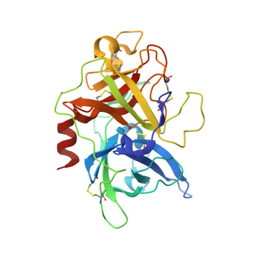
7VM4, 7VM5, 7VM6, 7VM7 - PubMed Abstract:
Nafamostat mesylate (NM) is a synthetic compound that inhibits various serine proteases produced during the coagulation cascade and inflammation. Previous studies showed that NM was a highly safe drug for the treatment of different cancers, but the precise functions and mechanisms of NM are not clear. In this study, we determined a series of crystal structures of NM and its hydrolysates in complex with a serine protease (urokinase-type plasminogen activator [uPA]). These structures reveal that NM was cleaved by uPA and that a hydrolyzed product (4-guanidinobenzoic acid [GBA]) remained covalently linked to Ser195 of uPA, and the other hydrolyzed product (6-amidino-2-naphthol [6A2N]) released from uPA. Strikingly, in the inactive uPA (uPA-S195A):NM structure, the 6A2N side of intact NM binds to the specific pocket of uPA. Molecular dynamics simulations and end-point binding free-energy calculations show that the conf1 of NM (6A2N as P1 group) in the uPA-S195A:NM complex may be more stable than conf2 of NM (GBA as P1 group). Moreover, in the structure of uPA:NM complex, the imidazole group of His57 flips further away from Ser195 and disrupts the stable canonical catalytic triad conformation. These results not only reveal the inhibitory mechanism of NM as an efficient serine protease inhibitor but also might provide the structural basis for the further development of serine protease inhibitors.
- College of Chemistry, Fuzhou University, Fuzhou, Fujian, P.R. China.
Organizational Affiliation: