7VLE

Oxy-deoxy intermediate of V2 hemoglobin at 55% oxygen saturation


Experimental Data Snapshot

  • Method: X-RAY DIFFRACTION
  • Resolution: 2.30 Å
  • R-Value Free: 0.246 
  • R-Value Work: 0.200 
  • R-Value Observed: 0.203 

wwPDB Validation   3D Report Full Report


Ligand Structure Quality Assessment 


This is version 1.2 of the entry. See complete history


Literature

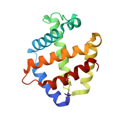
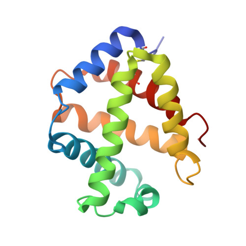
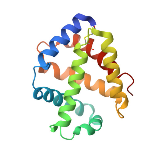
Structures of oxygen dissociation intermediates of 400 kDa V2 hemoglobin provide coarse snapshots of the protein allostery.

Numoto, N.Onoda, S.Kawano, Y.Okumura, H.Baba, S.Fukumori, Y.Miki, K.Ito, N.

(2022) Biophys Physicobiol 19: 1-10

  • DOI: https://doi.org/10.2142/biophysico.bppb-v19.0019
  • Primary Citation of Related Structures:  
    7VLC, 7VLD, 7VLE, 7VLF

  • PubMed Abstract: 

    Ever since the historic discovery of the cooperative oxygenation of its multiple subunits, hemoglobin (Hb) has been among the most exhaustively studied allosteric proteins. However, the lack of structural information on the intermediates between oxygenated and deoxygenated forms prevents our detailed understanding of the molecular mechanism of its allostery. It has been difficult to prepare crystals of intact oxy-deoxy intermediates and to individually identify the oxygen saturation for each subunit. However, our recent crystallographic studies have demonstrated that giant Hbs from annelids are suitable for overcoming these problems and can provide abundant information on oxy-deoxy intermediate structures. Here, we report the crystal structures of oxy-deoxy intermediates of a 400 kDa Hb (V2Hb) from the annelid Lamellibrachia satsuma , following up on a series of previous studies of similar giant Hbs. Four intermediate structures had average oxygen saturations of 78%, 69%, 55%, and 26%, as determined by the occupancy refinement of the bound oxygen based on ambient temperature factors. The structures demonstrate that the cooperative oxygen dissociation is weaker, large ternary and quaternary changes are induced at a later stage of the oxygen dissociation process, and the ternary and quaternary changes are smaller with local perturbations. Nonetheless, the overall structural transition seemed to proceed in the manner of the MWC two-state model. Our crystallographic snapshots of the allosteric transition of V2Hb provide important experimental evidence for a more detailed understanding of the allostery of Hbs by extension of the Monod-Wyman-Changeux (MWC) model.


  • Organizational Affiliation

    Medical Research Institute, Tokyo Medical and Dental University (TMDU), Bunkyo-ku, Tokyo 113-8510, Japan.


Macromolecules
Find similar proteins by:  (by identity cutoff)  |  3D Structure
Entity ID: 1
MoleculeChains Sequence LengthOrganismDetailsImage
Extracellular A1 globin
A, E
146Lamellibrachia satsumaMutation(s): 0 
UniProt
Find proteins for S0BBU7 (Lamellibrachia satsuma)
Explore S0BBU7 
Go to UniProtKB:  S0BBU7
Entity Groups  
Sequence Clusters30% Identity50% Identity70% Identity90% Identity95% Identity100% Identity
UniProt GroupS0BBU7
Sequence Annotations
Expand
  • Reference Sequence
Find similar proteins by:  (by identity cutoff)  |  3D Structure
Entity ID: 2
MoleculeChains Sequence LengthOrganismDetailsImage
Extracellular A2 globin
B, F
144Lamellibrachia satsumaMutation(s): 0 
UniProt
Find proteins for S0BBR6 (Lamellibrachia satsuma)
Explore S0BBR6 
Go to UniProtKB:  S0BBR6
Entity Groups  
Sequence Clusters30% Identity50% Identity70% Identity90% Identity95% Identity100% Identity
UniProt GroupS0BBR6
Sequence Annotations
Expand
  • Reference Sequence
Find similar proteins by:  (by identity cutoff)  |  3D Structure
Entity ID: 3
MoleculeChains Sequence LengthOrganismDetailsImage
Extracellular B2 globin
C, G
150Lamellibrachia satsumaMutation(s): 0 
UniProt
Find proteins for S0BCU7 (Lamellibrachia satsuma)
Explore S0BCU7 
Go to UniProtKB:  S0BCU7
Entity Groups  
Sequence Clusters30% Identity50% Identity70% Identity90% Identity95% Identity100% Identity
UniProt GroupS0BCU7
Sequence Annotations
Expand
  • Reference Sequence
Find similar proteins by:  (by identity cutoff)  |  3D Structure
Entity ID: 4
MoleculeChains Sequence LengthOrganismDetailsImage
Extracellular B1 globin
D, H
149Lamellibrachia satsumaMutation(s): 0 
UniProt
Find proteins for S0BAP9 (Lamellibrachia satsuma)
Explore S0BAP9 
Go to UniProtKB:  S0BAP9
Entity Groups  
Sequence Clusters30% Identity50% Identity70% Identity90% Identity95% Identity100% Identity
UniProt GroupS0BAP9
Sequence Annotations
Expand
  • Reference Sequence
Oligosaccharides

Help

Entity ID: 5
MoleculeChains Length2D Diagram Glycosylation3D Interactions
alpha-L-fucopyranose-(1-6)-2-acetamido-2-deoxy-beta-D-glucopyranose
I, J
2N-Glycosylation
Glycosylation Resources
GlyTouCan:  G86851RC
GlyCosmos:  G86851RC
GlyGen:  G86851RC
Small Molecules
Ligands 2 Unique
IDChains Name / Formula / InChI Key2D Diagram3D Interactions
HEM (Subject of Investigation/LOI)
Query on HEM

Download Ideal Coordinates CCD File 
K [auth A]
M [auth B]
O [auth C]
Q [auth D]
S [auth E]
K [auth A],
M [auth B],
O [auth C],
Q [auth D],
S [auth E],
U [auth F],
W [auth G],
Y [auth H]
PROTOPORPHYRIN IX CONTAINING FE
C34 H32 Fe N4 O4
KABFMIBPWCXCRK-RGGAHWMASA-L
OXY (Subject of Investigation/LOI)
Query on OXY

Download Ideal Coordinates CCD File 
L [auth A]
N [auth B]
P [auth C]
R [auth D]
T [auth E]
L [auth A],
N [auth B],
P [auth C],
R [auth D],
T [auth E],
V [auth F],
X [auth G],
Z [auth H]
OXYGEN MOLECULE
O2
MYMOFIZGZYHOMD-UHFFFAOYSA-N
Experimental Data & Validation

Experimental Data

  • Method: X-RAY DIFFRACTION
  • Resolution: 2.30 Å
  • R-Value Free: 0.246 
  • R-Value Work: 0.200 
  • R-Value Observed: 0.203 
  • Space Group: P 63
Unit Cell:
Length ( Å )Angle ( ˚ )
a = 109.644α = 90
b = 109.644β = 90
c = 195.496γ = 120
Software Package:
Software NamePurpose
PHENIXrefinement
XDSdata reduction
XDSdata scaling
PHASERphasing

Structure Validation

View Full Validation Report


Ligand Structure Quality Assessment 


Entry History & Funding Information

Deposition Data


Funding OrganizationLocationGrant Number
Japan Society for the Promotion of Science (JSPS)Japan25870200
Japan Society for the Promotion of Science (JSPS)Japan15K20971

Revision History  (Full details and data files)

  • Version 1.0: 2022-05-18
    Type: Initial release
  • Version 1.1: 2022-07-20
    Changes: Database references
  • Version 1.2: 2023-11-29
    Changes: Data collection, Refinement description