


The C-terminal DNA binding domain of EsrB from Edwardsiella piscicida
Liu, B., Reverter, D., Shao, S.To be published.
Experimental Data Snapshot
wwPDB Validation 3D Report Full Report

Entity ID: 1 | |||||
|---|---|---|---|---|---|
| Molecule | Chains | Sequence Length | Organism | Details | Image |
| Protein EsrB | 72 | Edwardsiella piscicida | Mutation(s): 0 Gene Names: EVK84_02890, MA13_contig00007-0127 | 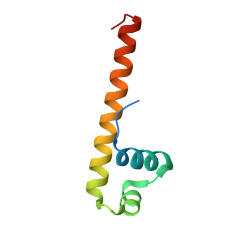 | |
| Length ( Å ) | Angle ( ˚ ) |
|---|---|
| a = 39.111 | α = 90 |
| b = 58.051 | β = 90 |
| c = 81.043 | γ = 90 |
| Software Name | Purpose |
|---|---|
| PHENIX | refinement |
| XSCALE | data scaling |
| PDB_EXTRACT | data extraction |
| XDS | data reduction |
| PHENIX | phasing |
| Funding Organization | Location | Grant Number |
|---|---|---|
| Spanish Ministry of Economy and Competitiveness | Spain | BFU2015-66417-P (MINECO/FEDER) |


