7VIH

Cryo-EM structure of Gi coupled Sphingosine 1-phosphate receptor bound with CBP-307


Experimental Data Snapshot

  • Method: ELECTRON MICROSCOPY
  • Resolution: 2.98 Å
  • Aggregation State: PARTICLE 
  • Reconstruction Method: SINGLE PARTICLE 

wwPDB Validation   3D Report Full Report


This is version 1.1 of the entry. See complete history


Literature

Structural insights into sphingosine-1-phosphate receptor activation.

Yu, L.He, L.Gan, B.Ti, R.Xiao, Q.Hu, H.Zhu, L.Wang, S.Ren, R.

(2022) Proc Natl Acad Sci U S A 119: e2117716119-e2117716119

  • DOI: https://doi.org/10.1073/pnas.2117716119
  • Primary Citation of Related Structures:  
    7VIE, 7VIF, 7VIG, 7VIH

  • PubMed Abstract: 

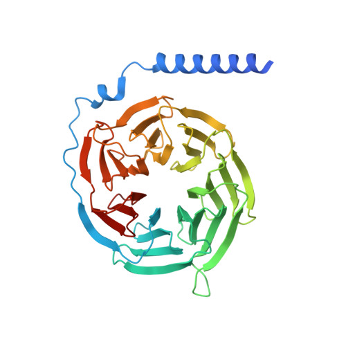
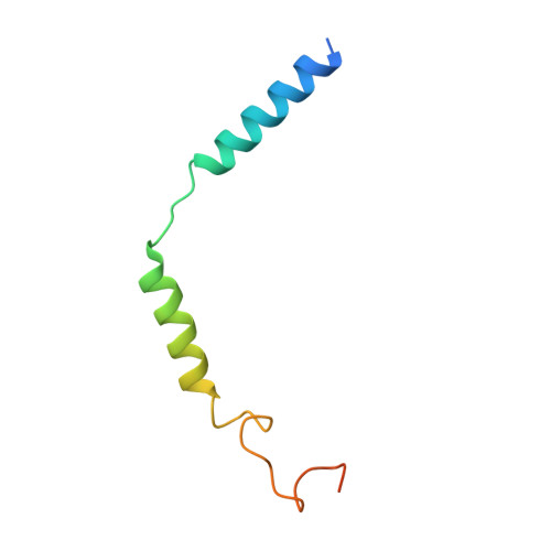
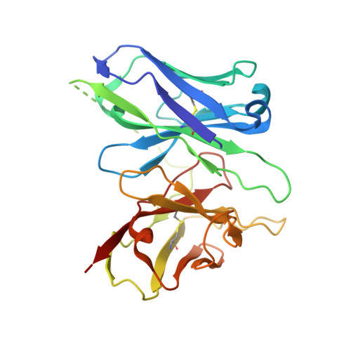
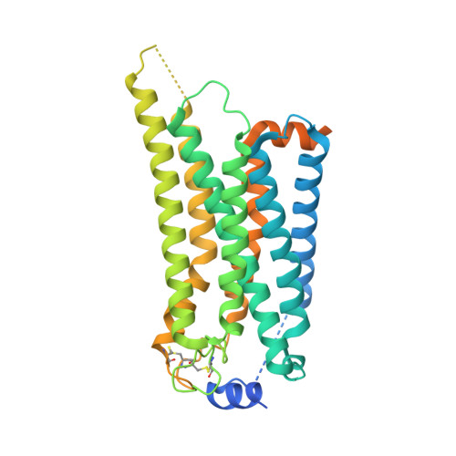
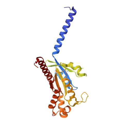
    As a critical sphingolipid metabolite, sphingosine-1-phosphate (S1P) plays an essential role in immune and vascular systems. There are five S1P receptors, designated as S1PR1 to S1PR5, encoded in the human genome, and their activities are governed by endogenous S1P, lipid-like S1P mimics, or nonlipid-like therapeutic molecules. Among S1PRs, S1PR1 stands out due to its nonredundant functions, such as the egress of T and B cells from the thymus and secondary lymphoid tissues, making it a potential therapeutic target. However, the structural basis of S1PR1 activation and regulation by various agonists remains unclear. Here, we report four atomic resolution cryo-electron microscopy (cryo-EM) structures of Gi-coupled human S1PR1 complexes: bound to endogenous agonist d18:1 S1P, benchmark lipid-like S1P mimic phosphorylated Fingolimod [(S)-FTY720-P], or nonlipid-like therapeutic molecule CBP-307 in two binding modes. Our results revealed the similarities and differences of activation of S1PR1 through distinct ligands binding to the amphiphilic orthosteric pocket. We also proposed a two-step “shallow to deep” transition process of CBP-307 for S1PR1 activation. Both binding modes of CBP-307 could activate S1PR1, but from shallow to deep transition may trigger the rotation of the N-terminal helix of Gαi and further stabilize the complex by increasing the Gαi interaction with the cell membrane. We combine with extensive biochemical analysis and molecular dynamic simulations to suggest key steps of S1P binding and receptor activation. The above results decipher the common feature of the S1PR1 agonist recognition and activation mechanism and will firmly promote the development of therapeutics targeting S1PRs.


  • Organizational Affiliation

    Kobilka Institute of Innovative Drug Discovery, School of Life and Health Sciences, The Chinese University of Hong Kong, Shenzhen 518172, China.


Macromolecules
Find similar proteins by:  (by identity cutoff)  |  3D Structure
Entity ID: 1
MoleculeChains Sequence LengthOrganismDetailsImage
Guanine nucleotide-binding protein G(I)/G(S)/G(T) subunit beta-1357Homo sapiensMutation(s): 0 
Gene Names: GNB1
UniProt & NIH Common Fund Data Resources
Find proteins for P62873 (Homo sapiens)
Explore P62873 
Go to UniProtKB:  P62873
PHAROS:  P62873
GTEx:  ENSG00000078369 
Entity Groups  
Sequence Clusters30% Identity50% Identity70% Identity90% Identity95% Identity100% Identity
UniProt GroupP62873
Sequence Annotations
Expand
  • Reference Sequence
Find similar proteins by:  (by identity cutoff)  |  3D Structure
Entity ID: 2
MoleculeChains Sequence LengthOrganismDetailsImage
Guanine nucleotide-binding protein G(I)/G(S)/G(O) subunit gamma-2B [auth C]71Homo sapiensMutation(s): 0 
Gene Names: GNG2
Membrane Entity: Yes 
UniProt & NIH Common Fund Data Resources
Find proteins for P59768 (Homo sapiens)
Explore P59768 
Go to UniProtKB:  P59768
PHAROS:  P59768
GTEx:  ENSG00000186469 
Entity Groups  
Sequence Clusters30% Identity50% Identity70% Identity90% Identity95% Identity100% Identity
UniProt GroupP59768
Sequence Annotations
Expand
  • Reference Sequence
Find similar proteins by:  (by identity cutoff)  |  3D Structure
Entity ID: 3
MoleculeChains Sequence LengthOrganismDetailsImage
scFv16C [auth E]251Mus musculusMutation(s): 0 
Entity Groups  
Sequence Clusters30% Identity50% Identity70% Identity90% Identity95% Identity100% Identity
Sequence Annotations
Expand
  • Reference Sequence
Find similar proteins by:  (by identity cutoff)  |  3D Structure
Entity ID: 4
MoleculeChains Sequence LengthOrganismDetailsImage
Sphingosine 1-phosphate receptor 1D [auth F]394Homo sapiensMutation(s): 0 
Gene Names: S1PR1CHEDG1EDG1
Membrane Entity: Yes 
UniProt & NIH Common Fund Data Resources
Find proteins for P21453 (Homo sapiens)
Explore P21453 
Go to UniProtKB:  P21453
PHAROS:  P21453
GTEx:  ENSG00000170989 
Entity Groups  
Sequence Clusters30% Identity50% Identity70% Identity90% Identity95% Identity100% Identity
UniProt GroupP21453
Sequence Annotations
Expand
  • Reference Sequence
Find similar proteins by:  (by identity cutoff)  |  3D Structure
Entity ID: 5
MoleculeChains Sequence LengthOrganismDetailsImage
Guanine nucleotide-binding protein G(i) subunit alpha-1E [auth D]354Homo sapiensMutation(s): 0 
Gene Names: GNAI1
Membrane Entity: Yes 
UniProt & NIH Common Fund Data Resources
Find proteins for P63096 (Homo sapiens)
Explore P63096 
Go to UniProtKB:  P63096
PHAROS:  P63096
GTEx:  ENSG00000127955 
Entity Groups  
Sequence Clusters30% Identity50% Identity70% Identity90% Identity95% Identity100% Identity
UniProt GroupP63096
Sequence Annotations
Expand
  • Reference Sequence
Small Molecules
Ligands 1 Unique
IDChains Name / Formula / InChI Key2D Diagram3D Interactions
7I4 (Subject of Investigation/LOI)
Query on 7I4

Download Ideal Coordinates CCD File 
F
1-[[2-fluoranyl-4-[5-[4-(2-methylpropyl)phenyl]-1,2,4-oxadiazol-3-yl]phenyl]methyl]azetidine-3-carboxylic acid
C23 H24 F N3 O3
YBIFMTGYWXNIRZ-UHFFFAOYSA-N
Experimental Data & Validation

Experimental Data

  • Method: ELECTRON MICROSCOPY
  • Resolution: 2.98 Å
  • Aggregation State: PARTICLE 
  • Reconstruction Method: SINGLE PARTICLE 

Structure Validation

View Full Validation Report


Entry History & Funding Information

Deposition Data


Funding OrganizationLocationGrant Number
National Natural Science Foundation of China (NSFC)China--

Revision History  (Full details and data files)

  • Version 1.0: 2022-09-28
    Type: Initial release
  • Version 1.1: 2022-10-05
    Changes: Database references