7VGW | pdb_00007vgw

Yeast gid10 with Pro-peptide


Experimental Data Snapshot

  • Method: X-RAY DIFFRACTION
  • Resolution: 2.80 Å
  • R-Value Free: 
    0.253 (Depositor), 0.250 (DCC) 
  • R-Value Work: 
    0.213 (Depositor), 0.210 (DCC) 
  • R-Value Observed: 
    0.217 (Depositor) 

Starting Model: experimental
View more details

wwPDB Validation   3D Report Full Report


This is version 1.1 of the entry. See complete history


Literature

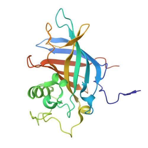
Crystal structure of yeast Gid10 in complex with Pro/N-degron.

Shin, J.S.Park, S.H.Kim, L.Heo, J.Song, H.K.

(2021) Biochem Biophys Res Commun 582: 86-92

  • DOI: https://doi.org/10.1016/j.bbrc.2021.10.007
  • Primary Citation of Related Structures:  
    7VGW

  • PubMed Abstract: 

    The cellular glucose level has to be tightly regulated by a variety of cellular processes. One of them is the degradation of gluconeogenic enzymes such as Fbp1, Icl1, Mdh2, and Pck1 by GID (glucose-induced degradation deficient) E3 ubiquitin ligase. The Gid4 component of the GID ligase complex is responsible for recognizing the N-terminal proline residue of the target substrates under normal conditions. However, an alternative N-recognin Gid10 controls the degradation process under stressed conditions. Although Gid10 shares a high sequence similarity with Gid4, their substrate specificities are quite different. Here, we report the structure of Gid10 from Saccharomyces cerevisiae in complex with Pro/N-degron, Pro-Tyr-Ile-Thr, which is almost identical to the sequence of the natural substrate Art2. Although Gid10 shares many structural features with the Gid4 protein from yeast and humans, the current structure explains the unique structural difference for the preference of bulky hydrophobic residue at the second position of Pro/N-degron. Therefore, this study provides a fundamental basis for understanding of the structural diversity and substrate specificity of recognition components in the GID E3 ligase complex involved in the Pro/N-degron pathway.


  • Organizational Affiliation
    • Department of Life Sciences, Korea University, 145 Anam-ro, Seongbuk-gu, Seoul, 02841, South Korea.

Macromolecules
Find similar proteins by:  (by identity cutoff)  |  3D Structure
Entity ID: 1
MoleculeChains Sequence LengthOrganismDetailsImage
BJ4_G0041530.mRNA.1.CDS.1
A, B
235Saccharomyces cerevisiaeMutation(s): 0 
Gene Names: PACBIOSEQ_LOCUS2470
UniProt
Find proteins for P53242 (Saccharomyces cerevisiae (strain ATCC 204508 / S288c))
Explore P53242 
Go to UniProtKB:  P53242
Entity Groups  
Sequence Clusters30% Identity50% Identity70% Identity90% Identity95% Identity100% Identity
UniProt GroupP53242
Sequence Annotations
Expand
  • Reference Sequence
Experimental Data & Validation

Experimental Data

  • Method: X-RAY DIFFRACTION
  • Resolution: 2.80 Å
  • R-Value Free:  0.253 (Depositor), 0.250 (DCC) 
  • R-Value Work:  0.213 (Depositor), 0.210 (DCC) 
  • R-Value Observed: 0.217 (Depositor) 
Space Group: C 2 2 21
Unit Cell:
Length ( Å )Angle ( ˚ )
a = 86.346α = 90
b = 109.913β = 90
c = 147.461γ = 90
Software Package:
Software NamePurpose
PHENIXrefinement
HKL-2000data reduction
PHENIXphasing

Structure Validation

View Full Validation Report


Entry History & Funding Information

Deposition Data


Funding OrganizationLocationGrant Number
National Research Foundation (NRF, Korea)Korea, Republic Of2021R1A6A3A01088067

Revision History  (Full details and data files)

  • Version 1.0: 2022-07-27
    Type: Initial release
  • Version 1.1: 2023-11-29
    Changes: Data collection, Refinement description