7VGT

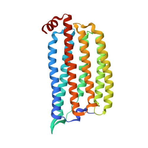
Time-resolved serial femtosecond crystallography structure of light-driven chloride ion-pumping rhodopsin, NM-R3: resting state structure with bromide ion


Experimental Data Snapshot

  • Method: X-RAY DIFFRACTION
  • Resolution: 2.10 Å
  • R-Value Free: 0.207 
  • R-Value Work: 0.176 
  • R-Value Observed: 0.177 

wwPDB Validation   3D Report Full Report


Ligand Structure Quality Assessment 


This is version 1.3 of the entry. See complete history


Literature

Conformational alterations in unidirectional ion transport of a light-driven chloride pump revealed using X-ray free electron lasers.

Hosaka, T.Nomura, T.Kubo, M.Nakane, T.Fangjia, L.Sekine, S.I.Ito, T.Murayama, K.Ihara, K.Ehara, H.Kashiwagi, K.Katsura, K.Akasaka, R.Hisano, T.Tanaka, T.Tanaka, R.Arima, T.Yamashita, A.Sugahara, M.Naitow, H.Matsuura, Y.Yoshizawa, S.Tono, K.Owada, S.Nureki, O.Kimura-Someya, T.Iwata, S.Nango, E.Shirouzu, M.

(2022) Proc Natl Acad Sci U S A 119

  • DOI: https://doi.org/10.1073/pnas.2117433119
  • Primary Citation of Related Structures:  
    7VGT, 7VGU, 7VGV

  • PubMed Abstract: 

    Light-driven chloride-pumping rhodopsins actively transport anions, including various halide ions, across cell membranes. Recent studies using time-resolved serial femtosecond crystallography (TR-SFX) have uncovered the structural changes and ion transfer mechanisms in light-driven cation-pumping rhodopsins. However, the mechanism by which the conformational changes pump an anion to achieve unidirectional ion transport, from the extracellular side to the cytoplasmic side, in anion-pumping rhodopsins remains enigmatic. We have collected TR-SFX data of Nonlabens marinus rhodopsin-3 (NM-R3), derived from a marine flavobacterium, at 10-µs and 1-ms time points after photoexcitation. Our structural analysis reveals the conformational alterations during ion transfer and after ion release. Movements of the retinal chromophore initially displace a conserved tryptophan to the cytoplasmic side of NM-R3, accompanied by a slight shift of the halide ion bound to the retinal. After ion release, the inward movements of helix C and helix G and the lateral displacements of the retinal block access to the extracellular side of NM-R3. Anomalous signal data have also been obtained from NM-R3 crystals containing iodide ions. The anomalous density maps provide insight into the halide binding site for ion transfer in NM-R3.


  • Organizational Affiliation

    RIKEN Center for Biosystems Dynamics Research, Yokohama, Kanagawa 230-0045, Japan.


Macromolecules
Find similar proteins by:  (by identity cutoff)  |  3D Structure
Entity ID: 1
MoleculeChains Sequence LengthOrganismDetailsImage
Chloride pumping rhodopsin279Nonlabens marinus S1-08Mutation(s): 0 
Gene Names: ClRNMS_1267
Membrane Entity: Yes 
UniProt
Find proteins for W8VZW3 (Nonlabens marinus S1-08)
Explore W8VZW3 
Go to UniProtKB:  W8VZW3
Entity Groups  
Sequence Clusters30% Identity50% Identity70% Identity90% Identity95% Identity100% Identity
UniProt GroupW8VZW3
Sequence Annotations
Expand
  • Reference Sequence
Small Molecules
Ligands 4 Unique
IDChains Name / Formula / InChI Key2D Diagram3D Interactions
RET
Query on RET

Download Ideal Coordinates CCD File 
B [auth A]RETINAL
C20 H28 O
NCYCYZXNIZJOKI-OVSJKPMPSA-N
D10
Query on D10

Download Ideal Coordinates CCD File 
G [auth A],
H [auth A],
I [auth A]
DECANE
C10 H22
DIOQZVSQGTUSAI-UHFFFAOYSA-N
HEX
Query on HEX

Download Ideal Coordinates CCD File 
C [auth A],
D [auth A],
E [auth A],
F [auth A]
HEXANE
C6 H14
VLKZOEOYAKHREP-UHFFFAOYSA-N
BR
Query on BR

Download Ideal Coordinates CCD File 
J [auth A],
K [auth A],
L [auth A]
BROMIDE ION
Br
CPELXLSAUQHCOX-UHFFFAOYSA-M
Experimental Data & Validation

Experimental Data

Unit Cell:
Length ( Å )Angle ( ˚ )
a = 104.3α = 90
b = 51.1β = 131.5
c = 78.4γ = 90
Software Package:
Software NamePurpose
PHENIXrefinement
PDB_EXTRACTdata extraction
CrystFELdata reduction
CrystFELdata scaling
PHENIXphasing

Structure Validation

View Full Validation Report


Ligand Structure Quality Assessment 


Entry History & Funding Information

Deposition Data


Funding OrganizationLocationGrant Number
Not funded--

Revision History  (Full details and data files)

  • Version 1.0: 2022-02-16
    Type: Initial release
  • Version 1.1: 2022-03-09
    Changes: Database references
  • Version 1.2: 2023-09-06
    Changes: Data collection, Database references
  • Version 1.3: 2023-11-29
    Changes: Refinement description