X-ray fluorescence holography of biological metal sites: Application to myoglobin.
Sato-Tomita, A., Ang, A.K.R., Kimura, K., Marumi, R., Happo, N., Matsushita, T., Park, S.Y., Shibayama, N., Sasaki, Y.C., Hayashi, K.(2022) Biochem Biophys Res Commun 635: 277-282
- PubMed: 36308907 Search on PubMed
- DOI: https://doi.org/10.1016/j.bbrc.2022.10.003
- Primary Citation Related Structures:
7VDN - PubMed Abstract:
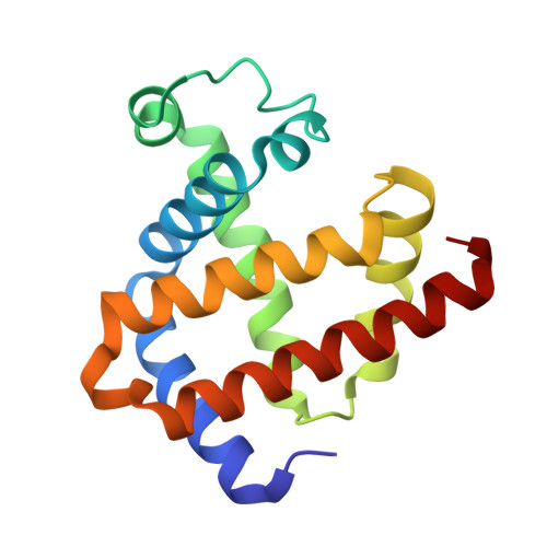
X-ray fluorescence holography (XFH) is a relatively new technique capable of providing unique three-dimensional structural information around specific atoms that act as a light source in crystalline samples. So far, XFH has typically been applied to inorganic materials such as dopants in metals and semiconductors. Here, we investigate the possibility of using XFH to visualize the metal active site in sperm whale myoglobin (Mb), a monomeric oxygen storage heme protein. We demonstrate that the atomic images reconstructed from the hologram data of crystals of carbonmonoxy myoglobin (MbCO) are moderately consistent with the crystal structure, which is also determined in this study by X-ray crystallography in the near-atomic resolution, as well as simulation results. These results open up a new avenue for the application of XFH to local atomic and electronic structure imaging of metal-sites in biomolecules.
- Division of Biophysics, Department of Physiology, Jichi Medical University, Yakushiji, Shimotsuke, Tochigi, 329-0498, Japan. Electronic address: ayana.sato@jichi.ac.jp.
Organizational Affiliation: