Discovery of Novel Drug-like PHGDH Inhibitors to Disrupt Serine Biosynthesis for Cancer Therapy.
Gao, D., Tang, S., Cen, Y., Yuan, L., Lan, X., Li, Q.H., Lin, G.Q., Huang, M., Tian, P.(2023) J Med Chem 66: 285-305
- PubMed: 36594670 Search on PubMed
- DOI: https://doi.org/10.1021/acs.jmedchem.2c01202
- Primary Citation Related Structures:
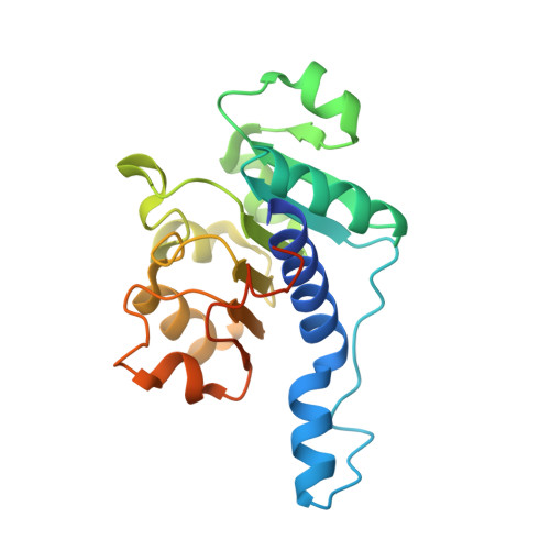
7VA1 - PubMed Abstract:
Being the rate-limiting enzyme within the serine biosynthesis pathway, phosphoglycerate dehydrogenase (PHGDH) is abnormally overexpressed in numerous malignant tumor cells and is a promising target for cancer treatment. Here, we report a series of novel PHGDH inhibitors using a focused compound screening and structural optimization approach. The lead compound D8 displayed good enzymatic inhibitory activity (IC 50 = 2.8 ± 0.1 μM), high binding affinity ( K d = 2.33 μM), and sensitivity to the cell lines with the PHGDH gene amplification or overexpression. Furthermore, D8 was proven to restrict the de novo serine synthesis from glucose within MDA-MB-468 cells. X-ray crystallographic analysis, molecular dynamics simulations, and mutagenesis experiments on PHGDH revealed the binding site at D175 inside the NAD + -binding pocket. Finally, D8 exhibited excellent in vivo pharmacokinetic properties ( F = 82.0%) and exerted evident antitumor efficacy in the PC9 xenograft mouse model.
- The Research Center of Chiral Drugs, Shanghai Frontiers Science Center for TCM Chemical Biology, Innovation Research Institute of Traditional Chinese Medicine, Shanghai University of Traditional Chinese Medicine, Shanghai 201203, China.
Organizational Affiliation: