Mechanistic insights into the subversion of the linear ubiquitin chain assembly complex by the E3 ligase IpaH1.4 of Shigella flexneri.
Liu, J., Wang, Y., Wang, D., Wang, Y., Xu, X., Zhang, Y., Li, Y., Zhang, M., Gong, X., Tang, Y., Shen, L., Li, M., Pan, L.(2022) Proc Natl Acad Sci U S A 119: e2116776119-e2116776119
- PubMed: 35294289 Search on PubMedSearch on PubMed Central
- DOI: https://doi.org/10.1073/pnas.2116776119
- Primary Citation Related Structures:
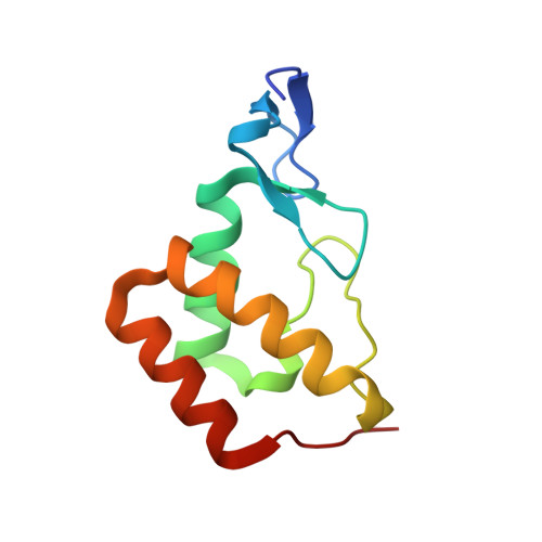
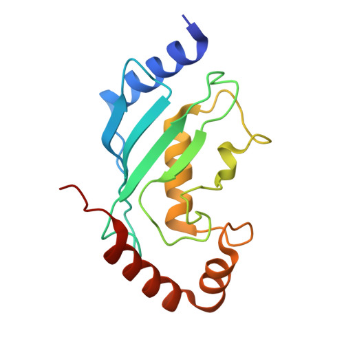
7V8E, 7V8F, 7V8G, 7V8H - PubMed Abstract:
Shigella flexneri, a gram-negative bacterium, is the major culprit of bacterial shigellosis and causes a large number of human infection cases and deaths worldwide annually. For evading the host immune response during infection, S. flexneri secrets two highly similar E3 ligases, IpaH1.4 and IpaH2.5, to subvert the linear ubiquitin chain assembly complex (LUBAC) of host cells, which is composed of HOIP, HOIL-1L, and SHARPIN. However, the detailed molecular mechanism underpinning the subversion of the LUBAC by IpaH1.4/2.5 remains elusive. Here, we demonstrated that IpaH1.4 can specifically recognize HOIP and HOIL-1L through its leucine-rich repeat (LRR) domain by binding to the HOIP RING1 domain and HOIL-1L ubiquitin-like (UBL) domain, respectively. The determined crystal structures of IpaH1.4 LRR/HOIP RING1, IpaH1.4 LRR/HOIL-1L UBL, and HOIP RING1/UBE2L3 complexes not only elucidate the binding mechanisms of IpaH1.4 with HOIP and HOIL-1L but also unveil that the recognition of HOIP by IpaH1.4 can inhibit the E2 binding of HOIP. Furthermore, we demonstrated that the interaction of IpaH1.4 LRR with HOIP RING1 or HOIL-1L UBL is essential for the ubiquitination of HOIP or HOIL-1L in vitro as well as the suppression of NF-κB activation by IpaH1.4 in cells. In summary, our work elucidated that in addition to inducing the proteasomal degradation of LUBAC, IpaH1.4 can also inhibit the E3 activity of LUBAC by blocking its E2 loading and/or disturbing its stability, thereby providing a paradigm showing how a bacterial E3 ligase adopts multiple tactics to subvert the key LUBAC of host cells.
- State Key Laboratory of Bioorganic and Natural Products Chemistry, Center for Excellence in Molecular Synthesis, Shanghai Institute of Organic Chemistry, University of Chinese Academy of Sciences, Chinese Academy of Sciences, Shanghai 200032, China.
Organizational Affiliation: