7V6B

Structure of the Dicer-2-R2D2 heterodimer


Experimental Data Snapshot

  • Method: ELECTRON MICROSCOPY
  • Resolution: 3.30 Å
  • Aggregation State: PARTICLE 
  • Reconstruction Method: SINGLE PARTICLE 

wwPDB Validation   3D Report Full Report


This is version 1.3 of the entry. See complete history


Literature

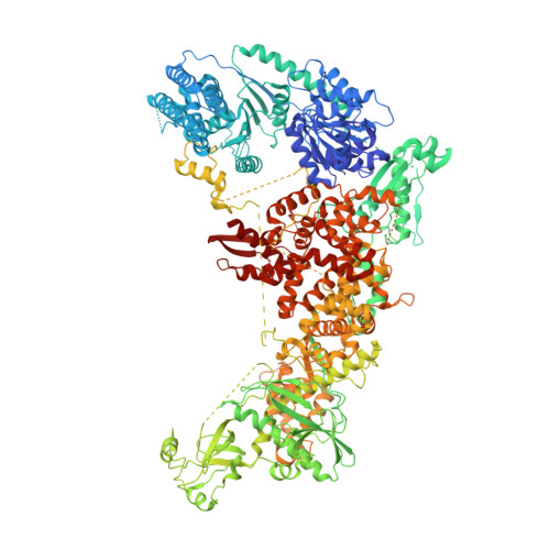
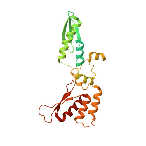
Structure of the Dicer-2-R2D2 heterodimer bound to a small RNA duplex.

Yamaguchi, S.Naganuma, M.Nishizawa, T.Kusakizako, T.Tomari, Y.Nishimasu, H.Nureki, O.

(2022) Nature 607: 393-398

  • DOI: https://doi.org/10.1038/s41586-022-04790-2
  • Primary Citation of Related Structures:  
    7V6B, 7V6C

  • PubMed Abstract: 

    In flies, Argonaute2 (Ago2) and small interfering RNA (siRNA) form an RNA-induced silencing complex to repress viral transcripts 1 . The RNase III enzyme Dicer-2 associates with its partner protein R2D2 and cleaves long double-stranded RNAs to produce 21-nucleotide siRNA duplexes, which are then loaded into Ago2 in a defined orientation 2-5 . Here we report cryo-electron microscopy structures of the Dicer-2-R2D2 and Dicer-2-R2D2-siRNA complexes. R2D2 interacts with the helicase domain and the central linker of Dicer-2 to inhibit the promiscuous processing of microRNA precursors by Dicer-2. Notably, our structure represents the strand-selection state in the siRNA-loading process, and reveals that R2D2 asymmetrically recognizes the end of the siRNA duplex with the higher base-pairing stability, and the other end is exposed to the solvent and is accessible by Ago2. Our findings explain how R2D2 senses the thermodynamic asymmetry of the siRNA and facilitates the siRNA loading into Ago2 in a defined orientation, thereby determining which strand of the siRNA duplex is used by Ago2 as the guide strand for target silencing.


  • Organizational Affiliation

    Department of Biological Sciences, Graduate School of Science, The University of Tokyo, Tokyo, Japan.


Macromolecules
Find similar proteins by:  (by identity cutoff)  |  3D Structure
Entity ID: 1
MoleculeChains Sequence LengthOrganismDetailsImage
Dicer-2, isoform A1,724Drosophila melanogasterMutation(s): 1 
Gene Names: 
EC: 3.1.21.1 (PDB Primary Data), 3.1.26 (PDB Primary Data), 3.1.26.3 (PDB Primary Data), 3.6.1.3 (PDB Primary Data)
UniProt
Find proteins for A1ZAW0 (Drosophila melanogaster)
Explore A1ZAW0 
Go to UniProtKB:  A1ZAW0
Entity Groups  
Sequence Clusters30% Identity50% Identity70% Identity90% Identity95% Identity100% Identity
UniProt GroupA1ZAW0
Sequence Annotations
Expand
  • Reference Sequence
Find similar proteins by:  (by identity cutoff)  |  3D Structure
Entity ID: 2
MoleculeChains Sequence LengthOrganismDetailsImage
R2D2315Drosophila melanogasterMutation(s): 0 
Gene Names: r2d2cg7138Dmel\CG7138R2D2Dmel_CG7138
UniProt
Find proteins for Q9VLW8 (Drosophila melanogaster)
Explore Q9VLW8 
Go to UniProtKB:  Q9VLW8
Entity Groups  
Sequence Clusters30% Identity50% Identity70% Identity90% Identity95% Identity100% Identity
UniProt GroupQ9VLW8
Sequence Annotations
Expand
  • Reference Sequence
Experimental Data & Validation

Experimental Data

  • Method: ELECTRON MICROSCOPY
  • Resolution: 3.30 Å
  • Aggregation State: PARTICLE 
  • Reconstruction Method: SINGLE PARTICLE 

Structure Validation

View Full Validation Report


Entry History & Funding Information

Deposition Data


Funding OrganizationLocationGrant Number
Japan Society for the Promotion of Science (JSPS)Japan202111067

Revision History  (Full details and data files)

  • Version 1.0: 2022-03-23
    Type: Initial release
  • Version 1.1: 2022-04-06
    Changes: Database references, Structure summary
  • Version 1.2: 2022-07-13
    Changes: Database references
  • Version 1.3: 2022-07-27
    Changes: Database references