


Mechanistic plasticity in ApmA enables aminoglycoside promiscuity for resistance
Bordeleau, E., Stogios, P.J., Evdokimova, E., Koteva, K., Savchenko, A., Wright, G.D.(2024) Nat Chem Biol 20: 234-242
Experimental Data Snapshot
Starting Model: experimental
View more details
(2024) Nat Chem Biol 20: 234-242
Entity ID: 1 | |||||
|---|---|---|---|---|---|
| Molecule | Chains | Sequence Length | Organism | Details | Image |
| Aminocyclitol acetyltransferase ApmA | 276 | Staphylococcus aureus | Mutation(s): 0 Gene Names: apmA | 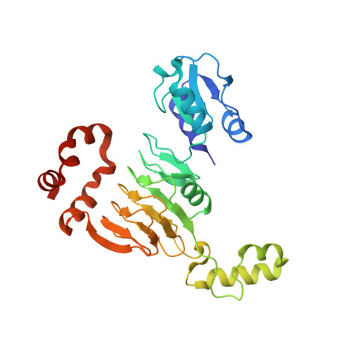 | |
UniProt | |||||
Entity Groups | |||||
| Sequence Clusters | 30% Identity50% Identity70% Identity90% Identity95% Identity100% Identity | ||||
| UniProt Group | A0A1D0AST6 | ||||
Sequence AnnotationsExpand | |||||
Reference Sequence | |||||
| Ligands 4 Unique | |||||
|---|---|---|---|---|---|
| ID | Chains | Name / Formula / InChI Key | 2D Diagram | 3D Interactions | |
| LLL (Subject of Investigation/LOI) Download:Ideal Coordinates CCD File | D [auth A], G [auth B], I [auth B] | (2R,3R,4R,5R)-2-((1S,2S,3R,4S,6R)-4,6-DIAMINO-3-((2R,3R,6S)-3-AMINO-6-(AMINOMETHYL)-TETRAHYDRO-2H-PYRAN-2-YLOXY)-2-HYDR
OXYCYCLOHEXYLOXY)-5-METHYL-4-(METHYLAMINO)-TETRAHYDRO-2H-PYRAN-3,5-DIOL C19 H39 N5 O7 VEGXETMJINRLTH-BOZYPMBZSA-N |  | ||
| EDO Download:Ideal Coordinates CCD File | E [auth A], H [auth B], K [auth C], M [auth C] | 1,2-ETHANEDIOL C2 H6 O2 LYCAIKOWRPUZTN-UHFFFAOYSA-N |  | ||
| CL Download:Ideal Coordinates CCD File | L [auth C] | CHLORIDE ION Cl VEXZGXHMUGYJMC-UHFFFAOYSA-M |  | ||
| MG Download:Ideal Coordinates CCD File | F [auth A], J [auth B] | MAGNESIUM ION Mg JLVVSXFLKOJNIY-UHFFFAOYSA-N |  | ||
| Length ( Å ) | Angle ( ˚ ) |
|---|---|
| a = 60.763 | α = 90 |
| b = 107.128 | β = 90 |
| c = 137.079 | γ = 90 |
| Software Name | Purpose |
|---|---|
| PHENIX | refinement |
| HKL-3000 | data reduction |
| HKL-3000 | data scaling |
| HKL-3000 | phasing |
| PHENIX | model building |
| Coot | model building |
| Funding Organization | Location | Grant Number |
|---|---|---|
| National Institutes of Health/National Institute Of Allergy and Infectious Diseases (NIH/NIAID) | United States | HHSN272201700060C |


