A Cell-Permeant Nanobody-Based Degrader That Induces Fetal Hemoglobin.
Shen, F., Zheng, G., Setegne, M., Tenglin, K., Izadi, M., Xie, H., Zhai, L., Orkin, S.H., Dassama, L.M.K.(2022) ACS Cent Sci 8: 1695-1703
- PubMed: 36589886 Search on PubMedSearch on PubMed Central
- DOI: https://doi.org/10.1021/acscentsci.2c00998
- Primary Citation Related Structures:
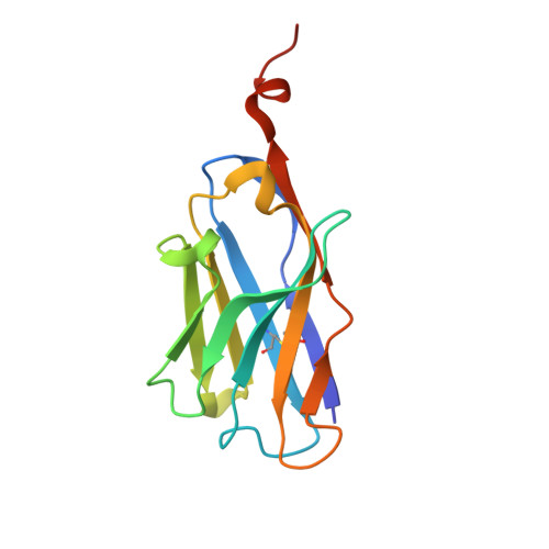
7UTG - PubMed Abstract:
Proximity-based strategies to degrade proteins have enormous therapeutic potential in medicine, but the technologies are limited to proteins for which small molecule ligands exist. The identification of such ligands for therapeutically relevant but "undruggable" proteins remains challenging. Herein, we employed yeast surface display of synthetic nanobodies to identify a protein ligand selective for BCL11A, a critical repressor of fetal globin gene transcription. Fusion of the nanobody to a cell-permeant miniature protein and an E3 adaptor creates a degrader that depletes cellular BCL11A in differentiated primary erythroid precursor cells, thereby inducing the expression of fetal hemoglobin, a modifier of clinical severity of sickle cell disease and β-thalassemia. Our strategy provides a means of fetal hemoglobin induction through reversible, temporal modulation of BCL11A. Additionally, it establishes a new paradigm for the targeted degradation of previously intractable proteins.
- Department of Chemistry and Sarafan ChEM-H, Stanford University, Stanford, California 94305, United States.
Organizational Affiliation: