7UTF | pdb_00007utf

Structure-Function characterization of an aldo-keto reductase involved in detoxification of the mycotoxin, deoxynivalenol


Experimental Data Snapshot

  • Method: X-RAY DIFFRACTION
  • Resolution: 2.50 Å
  • R-Value Free: 
    0.234 (Depositor), 0.233 (DCC) 
  • R-Value Work: 
    0.185 (Depositor), 0.185 (DCC) 
  • R-Value Observed: 
    0.187 (Depositor) 

Starting Model: experimental
View more details

wwPDB Validation 3D Report Full Report

Validation slider image for 7UTF

This is version 1.1 of the entry. See complete history

Literature

Structure-function characterization of an aldo-keto reductase involved in detoxification of the mycotoxin, deoxynivalenol.

Abraham, N.Schroeter, K.L.Zhu, Y.Chan, J.Evans, N.Kimber, M.S.Carere, J.Zhou, T.Seah, S.Y.K.

(2022) Sci Rep 12: 14737-14737

  • DOI: https://doi.org/10.1038/s41598-022-19040-8
  • Primary Citation Related Structures: 
    7UTF

  • PubMed Abstract: 

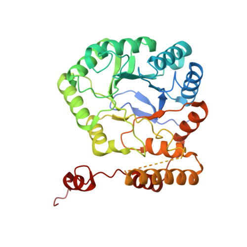
    Deoxynivalenol (DON) is a mycotoxin, produced by filamentous fungi such as Fusarium graminearum, that causes significant yield losses of cereal grain crops worldwide. One of the most promising methods to detoxify this mycotoxin involves its enzymatic epimerization to 3-epi-DON. DepB plays a critical role in this process by reducing 3-keto-DON, an intermediate in the epimerization process, to 3-epi-DON. DepB Rleg from Rhizobium leguminosarum is a member of the new aldo-keto reductase family, AKR18, and it has the unusual ability to utilize both NADH and NADPH as coenzymes, albeit with a 40-fold higher catalytic efficiency with NADPH compared to NADH. Structural analysis of DepB Rleg revealed the putative roles of Lys-217, Arg-290, and Gln-294 in NADPH specificity. Replacement of these residues by site-specific mutagenesis to negatively charged amino acids compromised NADPH binding with minimal effects on NADH binding. The substrate-binding site of DepB Rleg is larger than its closest structural homolog, AKR6A2, likely contributing to its ability to utilize a wide range of aldehydes and ketones, including the mycotoxin, patulin, as substrates. The structure of DepB Rleg also suggests that 3-keto-DON can adopt two binding modes to facilitate 4-pro-R hydride transfer to either the re- or si-face of the C3 ketone providing a possible explanation for the enzyme's ability to convert 3-keto-DON to 3-epi-DON and DON in diastereomeric ratios of 67.2% and 32.8% respectively.


  • Organizational Affiliation
    • Department of Molecular and Cellular Biology, University of Guelph, Guelph, Canada.

Macromolecule Content 

  • Total Structure Weight: 162.4 kDa 
  • Atom Count: 10,579 
  • Modeled Residue Count: 1,275 
  • Deposited Residue Count: 1,452 
  • Unique protein chains: 1

Macromolecules

Find similar proteins by:|  3D Structure
Entity ID: 1
MoleculeChains  Sequence LengthOrganismDetailsImage
Putative oxidoreductase, aryl-alcohol dehydrogenase like protein
A, B, C, D
363Rhizobium leguminosarumMutation(s): 0 
Gene Names: Rleg4DRAFT_7458
UniProt
Find proteins for J0WHR2 (Rhizobium leguminosarum bv. trifolii WSM2297)
Explore J0WHR2 
Go to UniProtKB:  J0WHR2
Entity Groups
Sequence Clusters30% Identity50% Identity70% Identity90% Identity95% Identity100% Identity
UniProt GroupJ0WHR2
Sequence Annotations
Expand
Reference Sequence

Small Molecules

Ligands 2 Unique
IDChains Name / Formula / InChI Key2D Diagram3D Interactions
FLC

Query on FLC


Download:Ideal Coordinates CCD File
M [auth A]CITRATE ANION
C6 H5 O7
KRKNYBCHXYNGOX-UHFFFAOYSA-K
SO4

Query on SO4


Download:Ideal Coordinates CCD File
AA [auth C]
BA [auth C]
CA [auth C]
DA [auth C]
E [auth A]
AA [auth C],
BA [auth C],
CA [auth C],
DA [auth C],
E [auth A],
EA [auth C],
F [auth A],
FA [auth D],
G [auth A],
GA [auth D],
H [auth A],
HA [auth D],
I [auth A],
IA [auth D],
J [auth A],
JA [auth D],
K [auth A],
KA [auth D],
L [auth A],
LA [auth D],
MA [auth D],
N [auth B],
O [auth B],
P [auth B],
Q [auth B],
R [auth B],
S [auth B],
T [auth B],
U [auth B],
V [auth B],
W [auth B],
X [auth C],
Y [auth C],
Z [auth C]
SULFATE ION
O4 S
QAOWNCQODCNURD-UHFFFAOYSA-L

Experimental Data & Validation

Experimental Data

  • Method: X-RAY DIFFRACTION
  • Resolution: 2.50 Å
  • R-Value Free:  0.234 (Depositor), 0.233 (DCC) 
  • R-Value Work:  0.185 (Depositor), 0.185 (DCC) 
  • R-Value Observed: 0.187 (Depositor) 
Space Group: P 4 21 2
Unit Cell:
Length ( Å )Angle ( ˚ )
a = 159.45α = 90
b = 159.45β = 90
c = 163.9γ = 90
Software Package:
Software NamePurpose
PHENIXrefinement
XDSdata reduction
XSCALEdata scaling
PHASERphasing

Structure Validation

View Full Validation Report


Entry History 

& Funding Information

Deposition Data


Funding OrganizationLocationGrant Number
Other privateCanadaJ-002433

Revision History  (Full details and data files)

  • Version 1.0: 2022-09-07
    Type: Initial release
  • Version 1.1: 2023-10-18
    Changes: Data collection, Refinement description