7UTC | pdb_00007utc

Crystal structure of secondary alcohol dehydrogenases from the Thermoanaerobacter ethanolicus with NADP and transition-state analogue inhibitor DMSO


Experimental Data Snapshot

  • Method: X-RAY DIFFRACTION
  • Resolution: 1.85 Å
  • R-Value Free: 
    0.227 (Depositor), 0.226 (DCC) 
  • R-Value Work: 
    0.178 (Depositor), 0.179 (DCC) 
  • R-Value Observed: 
    0.180 (Depositor) 

Starting Model: experimental
View more details

wwPDB Validation 3D Report Full Report

Validation slider image for 7UTC

Ligand Structure Quality Assessment 


This is version 1.4 of the entry. See complete history

Literature

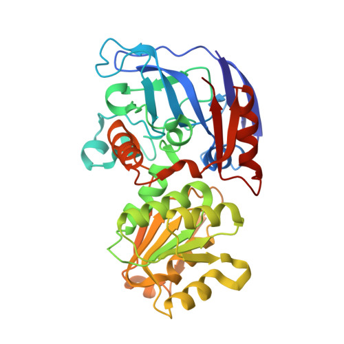
Crystallographic snapshots of ternary complexes of thermophilic secondary alcohol dehydrogenase from Thermoanaerobacter pseudoethanolicus reveal the dynamics of ligand exchange and the proton relay network.

Dinh, T.Rahn, K.T.Phillips, R.S.

(2022) Proteins 90: 1570-1583

  • DOI: https://doi.org/10.1002/prot.26339
  • Primary Citation Related Structures: 
    7UTC, 7UUT, 7UX4

  • PubMed Abstract: 

    Three-dimensional structures of I86A and C295A mutant secondary alcohol dehydrogenase (SADH) from Thermoanaerobacter pseudoethanolicus were determined by x-ray crystallography. The tetrameric structure of C295A-SADH soaked with NADP + and dimethyl sulfoxide (DMSO) was determined to 1.85 Å with an R free of 0.225. DMSO is bound to the tetrahedral zinc in each subunit, with ligands from SG of Cys-37, NE2 of His-59, and OD2 of Asp-150. The nicotinamide ring of NADP is hydrogen-bonded to the N of Ala-295 and the O of Val-265 and Gly-293. The O of DMSO is connected to a network of hydrogen bonds with OG of Ser-39, the 3'-OH of NADP, and ND1 of His-42. The structure of I86A-SADH soaked with 2-pentanol and NADP + contains (R)-2-pentanol bound in each subunit, ligated to the tetrahedral zinc, and connected to the proton relay network. The structure of I86A-SADH soaked with 3-methylcyclohexanol and NADP + has alcohol bound in three subunits. Two of the sites have the alcohol ligated to the zinc in an axial position, with OE2 of Glu-60 in the other axial position of a trigonal bipyramidal complex. One site has 3-methylcyclohexanol bound noncovalently, with the zinc in an inverted tetrahedral geometry with Glu-60. The fourth site also has the zinc in a trigonal bipyramidal complex with axial Glu-60 and water ligands. These structures demonstrate that ligand exchange of SADH involves pentacoordinate and inverted zinc complexes with Glu-60. Furthermore, we see a network of hydrogen bonds connecting the substrate oxygen to the external solvent that is likely to play a role in the mechanism of SADH.


  • Organizational Affiliation
    • Department of Chemistry, University of Georgia, Athens, Georgia, USA.

Macromolecule Content 

  • Total Structure Weight: 156.62 kDa 
  • Atom Count: 11,438 
  • Modeled Residue Count: 1,408 
  • Deposited Residue Count: 1,408 
  • Unique protein chains: 1

Macromolecules

Find similar proteins by:|  3D Structure
Entity ID: 1
MoleculeChains  Sequence LengthOrganismDetailsImage
Secondary-alcohol dehydrogenase
A, B, C, D
352Thermoanaerobacter pseudethanolicusMutation(s): 1 
EC: 1.1.1.80
UniProt
Find proteins for P14941 (Thermoanaerobacter brockii)
Explore P14941 
Go to UniProtKB:  P14941
Entity Groups
Sequence Clusters30% Identity50% Identity70% Identity90% Identity95% Identity100% Identity
UniProt GroupP14941
Sequence Annotations
Expand
Reference Sequence

Small Molecules

Ligands 3 Unique
IDChains Name / Formula / InChI Key2D Diagram3D Interactions
NAP
(Subject of Investigation/LOI)

Query on NAP


Download:Ideal Coordinates CCD File
AA [auth C],
F [auth A],
MA [auth D],
Q [auth B]
NADP NICOTINAMIDE-ADENINE-DINUCLEOTIDE PHOSPHATE
C21 H28 N7 O17 P3
XJLXINKUBYWONI-NNYOXOHSSA-N
DMS
(Subject of Investigation/LOI)

Query on DMS


Download:Ideal Coordinates CCD File
BA [auth C]
CA [auth C]
DA [auth C]
EA [auth C]
FA [auth C]
BA [auth C],
CA [auth C],
DA [auth C],
EA [auth C],
FA [auth C],
G [auth A],
GA [auth C],
H [auth A],
HA [auth C],
I [auth A],
IA [auth C],
J [auth A],
JA [auth C],
K [auth A],
KA [auth D],
L [auth A],
M [auth A],
N [auth A],
NA [auth D],
O [auth A],
OA [auth D],
PA [auth D],
QA [auth D],
R [auth B],
RA [auth D],
S [auth B],
SA [auth D],
T [auth B],
TA [auth D],
U [auth B],
UA [auth D],
V [auth B],
W [auth B],
X [auth B],
Y [auth B]
DIMETHYL SULFOXIDE
C2 H6 O S
IAZDPXIOMUYVGZ-UHFFFAOYSA-N
ZN
(Subject of Investigation/LOI)

Query on ZN


Download:Ideal Coordinates CCD File
E [auth A],
LA [auth D],
P [auth B],
Z [auth C]
ZINC ION
Zn
PTFCDOFLOPIGGS-UHFFFAOYSA-N

Experimental Data & Validation

Experimental Data

  • Method: X-RAY DIFFRACTION
  • Resolution: 1.85 Å
  • R-Value Free:  0.227 (Depositor), 0.226 (DCC) 
  • R-Value Work:  0.178 (Depositor), 0.179 (DCC) 
  • R-Value Observed: 0.180 (Depositor) 
Space Group: P 21 21 21
Unit Cell:
Length ( Å )Angle ( ˚ )
a = 79.709α = 90
b = 125.325β = 90
c = 137.366γ = 90
Software Package:
Software NamePurpose
PHENIXrefinement
XDSdata reduction
XSCALEdata scaling
PHASERphasing

Structure Validation

View Full Validation Report


Ligand Structure Quality Assessment 


Entry History 

& Funding Information

Deposition Data


Funding OrganizationLocationGrant Number
Not funded--

Revision History  (Full details and data files)

  • Version 1.0: 2022-05-04
    Type: Initial release
  • Version 1.1: 2022-05-11
    Changes: Database references
  • Version 1.2: 2022-05-18
    Changes: Database references, Source and taxonomy, Structure summary
  • Version 1.3: 2022-07-20
    Changes: Database references
  • Version 1.4: 2023-10-18
    Changes: Data collection, Refinement description