7UT3 | pdb_00007ut3

Crystal structure of complex of Fab, G10C with GalNAc-pNP

  • Classification: IMMUNE SYSTEM
  • Organism(s): Mus musculus
  • Expression System: Sapientia
  • Mutation(s): No 

  • Deposited: 2022-04-26 Released: 2022-09-21 
  • Deposition Author(s): Li, M., Wlodawer, A.
  • Funding Organization(s): National Institutes of Health/National Cancer Institute (NIH/NCI)

Experimental Data Snapshot

  • Method: X-RAY DIFFRACTION
  • Resolution: 3.00 Å
  • R-Value Free: 
    0.240 (Depositor), 0.231 (DCC) 
  • R-Value Work: 
    0.186 (Depositor), 0.187 (DCC) 
  • R-Value Observed: 
    0.188 (Depositor) 

Starting Models: experimental
View more details

wwPDB Validation 3D Report Full Report

Validation slider image for 7UT3

Ligand Structure Quality Assessment 


This is version 1.3 of the entry. See complete history

Literature

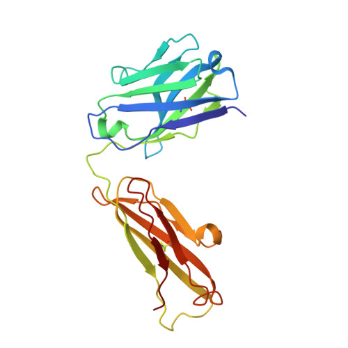
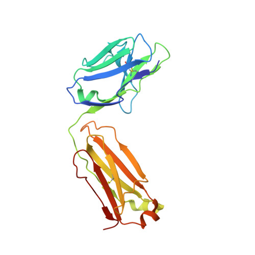
Development of a GalNAc-Tyrosine-Specific Monoclonal Antibody and Detection of Tyrosine O -GalNAcylation in Numerous Human Tissues and Cell Lines.

Xia, L.Bellomo, T.R.Gibadullin, R.Congdon, M.D.Edmondson, E.F.Li, M.Wlodawer, A.Li, C.Temme, J.S.Patel, P.Butcher, D.Gildersleeve, J.C.

(2022) J Am Chem Soc 144: 16410-16422

  • DOI: https://doi.org/10.1021/jacs.2c04477
  • Primary Citation Related Structures: 
    7UT3

  • PubMed Abstract: 

    Glycosylation is a vital post-translational modification involved in a range of biological processes including protein folding, signaling, and cell-cell interactions. In 2011, a new type of O -linked glycosylation was discovered, wherein the side-chain oxygen of tyrosine is modified with a GalNAc residue (GalNAc-Tyr). At present, very little is known about GalNAc-Tyr prevalence, function, or biosynthesis. Herein, we describe the design and synthesis of a GalNAc-Tyr-derived hapten and its use in generating a GalNAc-Tyr selective monoclonal antibody. The antibody, G10C, has an unusually high affinity (app K D = 100 pM) and excellent selectivity for GalNAc-Tyr. We also obtained a crystal structure of the G10C Fab region in complex with 4-nitrophenyl- N -acetyl-α-d-galactosaminide (a small molecule mimic of GalNAc-Tyr) providing insights into the structural basis for high affinity and selectivity. Using this antibody, we discovered that GalNAc-Tyr is widely expressed in most human tissues, indicating that it is a ubiquitous and underappreciated post-translational modification. Localization to specific cell types and organ substructures within those tissues indicates that GalNAc-Tyr is likely regulated in a cell-specific manner. GalNAc-Tyr was also observed in a variety of cell lines and primary cells but was only present on the external cell surface in certain cancer cell lines, suggesting that GalNAc-Tyr localization may be altered in cancer cells. Collectively, the results shed new light on this under-studied form of glycosylation and provide access to new tools that will enable expanded biochemical and clinical investigations.


  • Organizational Affiliation
    • Chemical Biology Laboratory, Center for Cancer Research, National Cancer Institute, Frederick, Maryland 21702, United States.

Macromolecule Content 

  • Total Structure Weight: 47.38 kDa 
  • Atom Count: 3,371 
  • Modeled Residue Count: 427 
  • Deposited Residue Count: 429 
  • Unique protein chains: 2

Macromolecules

Find similar proteins by:|  3D Structure
Entity ID: 1
MoleculeChains  Sequence LengthOrganismDetailsImage
Fab protein heavy chainA [auth H]217Mus musculusMutation(s): 0 
Find similar proteins by:|  3D Structure
Entity ID: 2
MoleculeChains  Sequence LengthOrganismDetailsImage
G10C Light chainB [auth L]212Mus musculusMutation(s): 0 

Small Molecules

Ligands 2 Unique
IDChains Name / Formula / InChI Key2D Diagram3D Interactions
TI0
(Subject of Investigation/LOI)

Query on TI0


Download:Ideal Coordinates CCD File
D [auth L]4-nitrophenyl 2-acetamido-2-deoxy-alpha-D-galactopyranoside
C14 H18 N2 O8
OMRLTNCLYHKQCK-RGDJUOJXSA-N
TRS

Query on TRS


Download:Ideal Coordinates CCD File
C [auth H]2-AMINO-2-HYDROXYMETHYL-PROPANE-1,3-DIOL
C4 H12 N O3
LENZDBCJOHFCAS-UHFFFAOYSA-O

Experimental Data & Validation

Experimental Data

  • Method: X-RAY DIFFRACTION
  • Resolution: 3.00 Å
  • R-Value Free:  0.240 (Depositor), 0.231 (DCC) 
  • R-Value Work:  0.186 (Depositor), 0.187 (DCC) 
  • R-Value Observed: 0.188 (Depositor) 
Space Group: P 61 2 2
Unit Cell:
Length ( Å )Angle ( ˚ )
a = 132.594α = 90
b = 132.594β = 90
c = 169.054γ = 120
Software Package:
Software NamePurpose
REFMACrefinement
PDB_EXTRACTdata extraction
HKL-2000data reduction
HKL-2000data scaling
PHASERphasing

Structure Validation

View Full Validation Report


Ligand Structure Quality Assessment 


Entry History 

& Funding Information

Deposition Data


Funding OrganizationLocationGrant Number
National Institutes of Health/National Cancer Institute (NIH/NCI)United States--

Revision History  (Full details and data files)

  • Version 1.0: 2022-09-21
    Type: Initial release
  • Version 1.1: 2022-09-28
    Changes: Database references
  • Version 1.2: 2023-10-18
    Changes: Data collection, Refinement description
  • Version 1.3: 2024-10-23
    Changes: Structure summary