7UPZ

Structural basis for cell type specific DNA binding of C/EBPbeta: the case of cell cycle inhibitor p15INK4b promoter


Experimental Data Snapshot

  • Method: X-RAY DIFFRACTION
  • Resolution: 2.49 Å
  • R-Value Free: 0.264 
  • R-Value Work: 0.212 
  • R-Value Observed: 0.215 

wwPDB Validation   3D Report Full Report


This is version 1.1 of the entry. See complete history


Literature

Structural basis for cell type specific DNA binding of C/EBP beta : The case of cell cycle inhibitor p15INK4b promoter.

Lountos, G.T.Cherry, S.Tropea, J.E.Wlodawer, A.Miller, M.

(2022) J Struct Biol 214: 107918-107918

  • DOI: https://doi.org/10.1016/j.jsb.2022.107918
  • Primary Citation of Related Structures:  
    7UPZ

  • PubMed Abstract: 

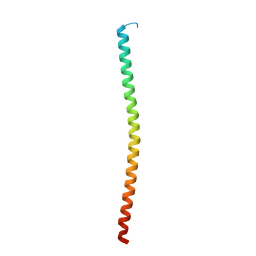
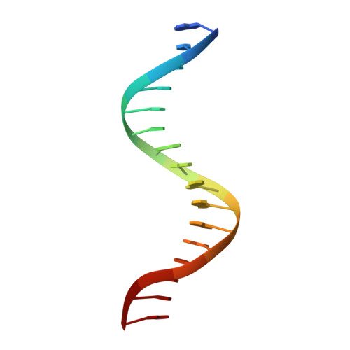
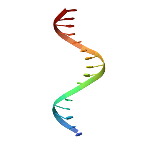
    C/EBPβ is a key regulator of numerous cellular processes, but it can also contribute to tumorigenesis and viral diseases. It binds to specific DNA sequences (C/EBP sites) and interacts with other transcription factors to control expression of multiple eukaryotic genes in a tissue and cell-type dependent manner. A body of evidence has established that cell-type-specific regulatory information is contained in the local DNA sequence of the binding motif. In human epithelial cells, C/EBPβ is an essential cofactor for TGFβ signaling in the case of Smad2/3/4 and FoxO-dependent induction of the cell cycle inhibitor, p15INK4b. In the TGFβ-responsive region 2 of the p15INK4b promoter, the Smad binding site is flanked by a C/EBP site, CTTAA•GAAAG, which differs from the canonical, palindromic ATTGC•GCAAT motif. The X-ray crystal structure of C/EBPβ bound to the p15INK4b promoter fragment shows how GCGC-to-AAGA substitution generates changes in the intermolecular interactions in the protein-DNA interface that enhances C/EBPβ binding specificity, limits possible epigenetic regulation of the promoter, and generates a DNA element with a unique pattern of methyl groups in the major groove. Significantly, CT/GA dinucleotides located at the 5'ends of the double stranded element maintain local narrowing of the DNA minor groove width that is necessary for DNA recognition. Our results suggest that C/EBPβ would accept all forms of modified cytosine in the context of the CpT site. This contrasts with the effect on the consensus motif, where C/EBPβ binding is modestly increased by cytosine methylation, but substantially decreased by hydroxymethylation.


  • Organizational Affiliation

    Basic Science Program, Frederick National Laboratory for Cancer Research, Frederick, MD 21702, USA. Electronic address: lountosg@mail.nih.gov.


Macromolecules

Find similar proteins by:  (by identity cutoff)  |  3D Structure
Entity ID: 1
MoleculeChains Sequence LengthOrganismDetailsImage
CCAAT/enhancer-binding protein beta
A, B
80Homo sapiensMutation(s): 0 
Gene Names: CEBPBTCF5PP9092
UniProt & NIH Common Fund Data Resources
Find proteins for P17676 (Homo sapiens)
Explore P17676 
Go to UniProtKB:  P17676
PHAROS:  P17676
GTEx:  ENSG00000172216 
Entity Groups  
Sequence Clusters30% Identity50% Identity70% Identity90% Identity95% Identity100% Identity
UniProt GroupP17676
Sequence Annotations
Expand
  • Reference Sequence

Find similar nucleic acids by:  Sequence   |   3D Structure  

Entity ID: 2
MoleculeChains LengthOrganismImage
DNA (5'-D(*AP*TP*TP*CP*TP*TP*AP*AP*GP*AP*AP*AP*GP*AP*CP*G)-3')16Homo sapiens
Sequence Annotations
Expand
  • Reference Sequence

Find similar nucleic acids by:  Sequence   |   3D Structure  

Entity ID: 3
MoleculeChains LengthOrganismImage
DNA (5'-D(*TP*CP*GP*TP*CP*TP*TP*TP*CP*TP*TP*AP*AP*GP*AP*A)-3')16Homo sapiens
Sequence Annotations
Expand
  • Reference Sequence
Experimental Data & Validation

Experimental Data

  • Method: X-RAY DIFFRACTION
  • Resolution: 2.49 Å
  • R-Value Free: 0.264 
  • R-Value Work: 0.212 
  • R-Value Observed: 0.215 
  • Space Group: C 2 2 21
Unit Cell:
Length ( Å )Angle ( ˚ )
a = 101.699α = 90
b = 112.758β = 90
c = 75.528γ = 90
Software Package:
Software NamePurpose
PHENIXrefinement
PDB_EXTRACTdata extraction
HKL-3000data reduction
HKL-3000data scaling
PHASERphasing

Structure Validation

View Full Validation Report


Entry History & Funding Information

Deposition Data


Funding OrganizationLocationGrant Number
National Institutes of Health/National Cancer Institute (NIH/NCI)United States--

Revision History  (Full details and data files)

  • Version 1.0: 2022-11-23
    Type: Initial release
  • Version 1.1: 2023-10-18
    Changes: Data collection, Refinement description