7UN7

DNA Polymerase lambda in complex with a 1nt microhomology substrate

  • Classification: REPLICATION
  • Organism(s): Homo sapiens
  • Expression System: Escherichia coli
  • Mutation(s): Yes 

  • Deposited: 2022-04-09 Released: 2023-03-29 
  • Deposition Author(s): Jamsen, J.A.
  • Funding Organization(s): National Institutes of Health/National Institute of Environmental Health Sciences (NIH/NIEHS)

Experimental Data Snapshot

  • Method: X-RAY DIFFRACTION
  • Resolution: 2.04 Å
  • R-Value Free: 0.247 
  • R-Value Work: 0.220 
  • R-Value Observed: 0.221 

wwPDB Validation   3D Report Full Report


Ligand Structure Quality Assessment 


This is version 1.1 of the entry. See complete history


Literature

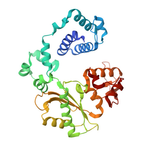
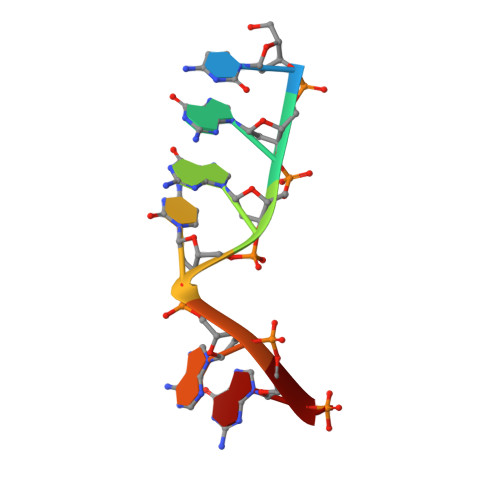
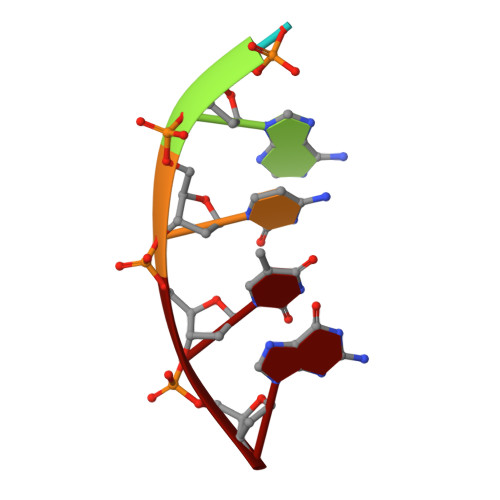
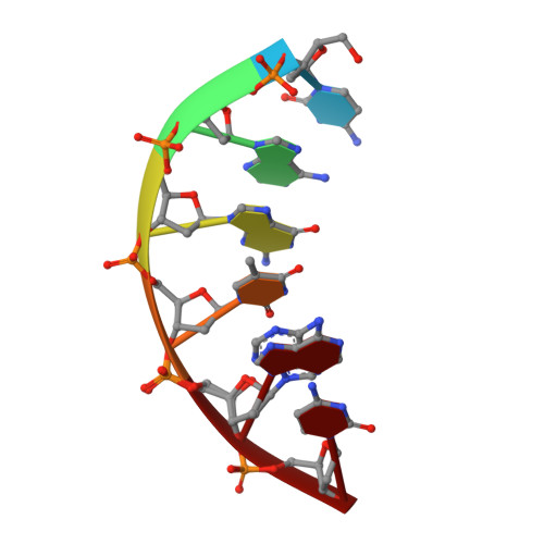
Pol lambda promotes microhomology-mediated end-joining.

Chandramouly, G.Jamsen, J.Borisonnik, N.Tyagi, M.Calbert, M.L.Tredinnick, T.Ozdemir, A.Y.Kent, T.Demidova, E.V.Arora, S.Wilson, S.H.Pomerantz, R.T.

(2023) Nat Struct Mol Biol 30: 107-114

  • DOI: https://doi.org/10.1038/s41594-022-00895-4
  • Primary Citation of Related Structures:  
    7UN7

  • PubMed Abstract: 

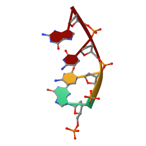
    The double-strand break (DSB) repair pathway called microhomology-mediated end-joining (MMEJ) is thought to be dependent on DNA polymerase theta (Polθ) and occur independently of nonhomologous end-joining (NHEJ) factors. An unresolved question is whether MMEJ is facilitated by a single Polθ-mediated end-joining pathway or consists of additional undiscovered pathways. We find that human X-family Polλ, which functions in NHEJ, additionally exhibits robust MMEJ activity like Polθ. Polλ promotes MMEJ in mammalian cells independently of essential NHEJ factors LIG4/XRCC4 and Polθ, which reveals a distinct Polλ-dependent MMEJ mechanism. X-ray crystallography employing in situ photo-induced DSB formation captured Polλ in the act of stabilizing a microhomology-mediated DNA synapse with incoming nucleotide at 2.0 Å resolution and reveals how Polλ performs replication across a DNA synapse joined by minimal base-pairing. Last, we find that Polλ is semisynthetic lethal with BRCA1 and BRCA2. Together, these studies indicate Polλ MMEJ as a distinct DSB repair mechanism.


  • Organizational Affiliation

    Department of Biochemistry and Molecular Biology, Sidney Kimmel Cancer Center, Thomas Jefferson University, Philadelphia, PA, USA.


Macromolecules

Find similar proteins by:  (by identity cutoff)  |  3D Structure
Entity ID: 1
MoleculeChains Sequence LengthOrganismDetailsImage
DNA polymerase lambda329Homo sapiensMutation(s): 1 
Gene Names: POLL
EC: 2.7.7.7 (PDB Primary Data), 4.2.99 (PDB Primary Data)
UniProt & NIH Common Fund Data Resources
Find proteins for Q9UGP5 (Homo sapiens)
Explore Q9UGP5 
Go to UniProtKB:  Q9UGP5
PHAROS:  Q9UGP5
GTEx:  ENSG00000166169 
Entity Groups  
Sequence Clusters30% Identity50% Identity70% Identity90% Identity95% Identity100% Identity
UniProt GroupQ9UGP5
Sequence Annotations
Expand
  • Reference Sequence

Find similar nucleic acids by:  Sequence   |   3D Structure  

Entity ID: 2
MoleculeChains LengthOrganismImage
DNA (5'-D(*CP*GP*GP*CP*AP*GP*T)-3')B [auth T]7Homo sapiens
Sequence Annotations
Expand
  • Reference Sequence

Find similar nucleic acids by:  Sequence   |   3D Structure  

Entity ID: 3
MoleculeChains LengthOrganismImage
DNA (5'-D(*TP*AP*CP*TP*G)-3')C [auth U]5Homo sapiens
Sequence Annotations
Expand
  • Reference Sequence

Find similar nucleic acids by:  Sequence   |   3D Structure  

Entity ID: 4
MoleculeChains LengthOrganismImage
DNA (5'-D(*CP*AP*GP*TP*AP*C)-3')D [auth P]6Homo sapiens
Sequence Annotations
Expand
  • Reference Sequence

Find similar nucleic acids by:  Sequence   |   3D Structure  

Entity ID: 5
MoleculeChains LengthOrganismImage
DNA (5'-D(P*GP*CP*CP*G)-3')E [auth D]4Homo sapiens
Sequence Annotations
Expand
  • Reference Sequence
Small Molecules
Ligands 5 Unique
IDChains Name / Formula / InChI Key2D Diagram3D Interactions
TTP (Subject of Investigation/LOI)
Query on TTP

Download Ideal Coordinates CCD File 
F [auth A]THYMIDINE-5'-TRIPHOSPHATE
C10 H17 N2 O14 P3
NHVNXKFIZYSCEB-XLPZGREQSA-N
GOL
Query on GOL

Download Ideal Coordinates CCD File 
G [auth A]GLYCEROL
C3 H8 O3
PEDCQBHIVMGVHV-UHFFFAOYSA-N
EDO
Query on EDO

Download Ideal Coordinates CCD File 
L [auth A],
M [auth A],
N [auth A],
O [auth A]
1,2-ETHANEDIOL
C2 H6 O2
LYCAIKOWRPUZTN-UHFFFAOYSA-N
CA (Subject of Investigation/LOI)
Query on CA

Download Ideal Coordinates CCD File 
J [auth A],
K [auth A]
CALCIUM ION
Ca
BHPQYMZQTOCNFJ-UHFFFAOYSA-N
NA
Query on NA

Download Ideal Coordinates CCD File 
H [auth A],
I [auth A]
SODIUM ION
Na
FKNQFGJONOIPTF-UHFFFAOYSA-N
Experimental Data & Validation

Experimental Data

  • Method: X-RAY DIFFRACTION
  • Resolution: 2.04 Å
  • R-Value Free: 0.247 
  • R-Value Work: 0.220 
  • R-Value Observed: 0.221 
  • Space Group: P 21 21 21
Unit Cell:
Length ( Å )Angle ( ˚ )
a = 56.135α = 90
b = 62.774β = 90
c = 140.193γ = 90
Software Package:
Software NamePurpose
PHENIXrefinement
autoPROCdata scaling
PDB_EXTRACTdata extraction
autoPROCdata reduction
PHENIXphasing

Structure Validation

View Full Validation Report


Ligand Structure Quality Assessment 


Entry History & Funding Information

Deposition Data

  • Released Date: 2023-03-29 
  • Deposition Author(s): Jamsen, J.A.

Funding OrganizationLocationGrant Number
National Institutes of Health/National Institute of Environmental Health Sciences (NIH/NIEHS)United StatesK99ES029572-01
National Institutes of Health/National Institute of Environmental Health Sciences (NIH/NIEHS)United States4R00ES029572 - 02

Revision History  (Full details and data files)

  • Version 1.0: 2023-03-29
    Type: Initial release
  • Version 1.1: 2023-10-25
    Changes: Data collection, Refinement description