7UMB

NanoBRET tracer Tram-bo bound to a KSR2-MEK1 complex


Experimental Data Snapshot

  • Method: X-RAY DIFFRACTION
  • Resolution: 3.23 Å
  • R-Value Free: 0.268 
  • R-Value Work: 0.229 
  • R-Value Observed: 0.231 

wwPDB Validation   3D Report Full Report


Ligand Structure Quality Assessment 


This is version 1.2 of the entry. See complete history


Literature

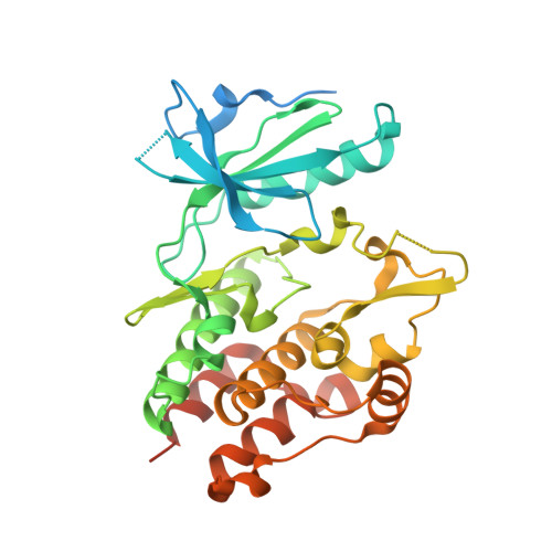
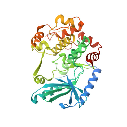
Live-cell target engagement of allosteric MEKi on MEK-RAF/KSR-14-3-3 complexes.

Marsiglia, W.M.Chow, A.Khan, Z.M.He, L.Dar, A.C.

(2024) Nat Chem Biol 20: 373-381

  • DOI: https://doi.org/10.1038/s41589-023-01454-8
  • Primary Citation of Related Structures:  
    7UMB

  • PubMed Abstract: 

    The RAS-mitogen-activated protein kinase (MAPK) pathway includes KSR, RAF, MEK and the phospho-regulatory sensor 14-3-3. Specific assemblies among these components drive various diseases and likely dictate efficacy for numerous targeted therapies, including allosteric MEK inhibitors (MEKi). However, directly measuring drug interactions on physiological RAS-MAPK complexes in live cells has been inherently challenging to query and therefore remains poorly understood. Here we present a series of NanoBRET-based assays to quantify direct target engagement of MEKi on MEK1 and higher-order MEK1-bound complexes with ARAF, BRAF, CRAF, KSR1 and KSR2 in the presence and absence of 14-3-3 in living cells. We find distinct MEKi preferences among these complexes that can be compiled to generate inhibitor binding profiles. Further, these assays can report on the influence of the pathogenic BRAF-V600E mutant on MEKi binding. Taken together, these approaches can be used as a platform to screen for compounds intended to target specific complexes in the RAS-MAPK cascade.


  • Organizational Affiliation

    Department of Oncological Sciences, The Tisch Cancer Institute, Mount Sinai Center for Therapeutic Discovery, Icahn School of Medicine at Mount Sinai, New York, NY, USA. wmmarsig@uab.edu.


Macromolecules
Find similar proteins by:  (by identity cutoff)  |  3D Structure
Entity ID: 1
MoleculeChains Sequence LengthOrganismDetailsImage
Kinase suppressor of Ras 2A [auth B]342Homo sapiensMutation(s): 0 
Gene Names: KSR2
EC: 2.7.11.1
UniProt & NIH Common Fund Data Resources
Find proteins for Q6VAB6 (Homo sapiens)
Explore Q6VAB6 
Go to UniProtKB:  Q6VAB6
PHAROS:  Q6VAB6
GTEx:  ENSG00000171435 
Entity Groups  
Sequence Clusters30% Identity50% Identity70% Identity90% Identity95% Identity100% Identity
UniProt GroupQ6VAB6
Sequence Annotations
Expand
  • Reference Sequence
Find similar proteins by:  (by identity cutoff)  |  3D Structure
Entity ID: 2
MoleculeChains Sequence LengthOrganismDetailsImage
Dual specificity mitogen-activated protein kinase kinase 1B [auth C]384Homo sapiensMutation(s): 0 
Gene Names: MAP2K1
UniProt & NIH Common Fund Data Resources
Find proteins for Q02750 (Homo sapiens)
Explore Q02750 
Go to UniProtKB:  Q02750
PHAROS:  Q02750
GTEx:  ENSG00000169032 
Entity Groups  
Sequence Clusters30% Identity50% Identity70% Identity90% Identity95% Identity100% Identity
UniProt GroupQ02750
Sequence Annotations
Expand
  • Reference Sequence
Small Molecules
Ligands 2 Unique
IDChains Name / Formula / InChI Key2D Diagram3D Interactions
U9F (Subject of Investigation/LOI)
Query on U9F

Download Ideal Coordinates CCD File 
E [auth C]{3-[(1M,2M)-2-{[(1S,2P)-1H,1'H-[2,2'-bipyrrol]-5-yl-kappaN~1~]methylidene}-2H-pyrrol-5-yl-kappaN]-N-(2-{2-[3-({(3M)-3-[(4aM)-3-cyclopropyl-5-(2-fluoro-4-iodoanilino)-6,8-dimethyl-2,4,7-trioxo-3,4,6,7-tetrahydropyrido[4,3-d]pyrimidin-1(2H)-yl]phenyl}amino)-3-oxopropoxy]ethoxy}ethyl)propanamidato}(difluorido)boron
C47 H46 B F3 I N9 O7
IBAMXDPOERGEKA-UHFFFAOYSA-N
ANP
Query on ANP

Download Ideal Coordinates CCD File 
C [auth B],
D [auth C]
PHOSPHOAMINOPHOSPHONIC ACID-ADENYLATE ESTER
C10 H17 N6 O12 P3
PVKSNHVPLWYQGJ-KQYNXXCUSA-N
Experimental Data & Validation

Experimental Data

  • Method: X-RAY DIFFRACTION
  • Resolution: 3.23 Å
  • R-Value Free: 0.268 
  • R-Value Work: 0.229 
  • R-Value Observed: 0.231 
  • Space Group: P 61 2 2
Unit Cell:
Length ( Å )Angle ( ˚ )
a = 139.5α = 90
b = 139.5β = 90
c = 220γ = 120
Software Package:
Software NamePurpose
PHENIXrefinement
XDSdata reduction
XDSdata scaling
PHASERphasing

Structure Validation

View Full Validation Report


Ligand Structure Quality Assessment 


Entry History & Funding Information

Deposition Data


Funding OrganizationLocationGrant Number
National Institutes of Health/National Cancer Institute (NIH/NCI)United States--

Revision History  (Full details and data files)

  • Version 1.0: 2023-09-27
    Type: Initial release
  • Version 1.1: 2023-11-22
    Changes: Database references
  • Version 1.2: 2024-03-20
    Changes: Database references