Phosphorylation of SAMHD1 Thr592 increases C-terminal domain dynamics, tetramer dissociation and ssDNA binding kinetics.
Orris, B., Huynh, K.W., Ammirati, M., Han, S., Bolanos, B., Carmody, J., Petroski, M.D., Bosbach, B., Shields, D.J., Stivers, J.T.(2022) Nucleic Acids Res 50: 7545-7559
- PubMed: 35801923 Search on PubMedSearch on PubMed Central
- DOI: https://doi.org/10.1093/nar/gkac573
- Primary Citation Related Structures:
7UJN - PubMed Abstract:
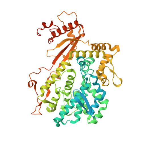
SAM and HD domain containing deoxynucleoside triphosphate triphosphohydrolase 1 (SAMHD1) is driven into its activated tetramer form by binding of GTP activator and dNTP activators/substrates. In addition, the inactive monomeric and dimeric forms of the enzyme bind to single-stranded (ss) nucleic acids. During DNA replication SAMHD1 can be phosphorylated by CDK1 and CDK2 at its C-terminal threonine 592 (pSAMHD1), localizing the enzyme to stalled replication forks (RFs) to promote their restart. Although phosphorylation has only a small effect on the dNTPase activity and ssDNA binding affinity of SAMHD1, perturbation of the native T592 by phosphorylation decreased the thermal stability of tetrameric SAMHD1 and accelerated tetramer dissociation in the absence and presence of ssDNA (∼15-fold). In addition, we found that ssDNA binds competitively with GTP to the A1 site. A full-length SAMHD1 cryo-EM structure revealed substantial dynamics in the C-terminal domain (which contains T592), which could be modulated by phosphorylation. We propose that T592 phosphorylation increases tetramer dynamics and allows invasion of ssDNA into the A1 site and the previously characterized DNA binding surface at the dimer-dimer interface. These features are consistent with rapid and regiospecific inactivation of pSAMHD1 dNTPase at RFs or other sites of free ssDNA in cells.
- Department of Pharmacology and Molecular Sciences, Johns Hopkins University School of Medicine 725 North Wolfe Street Baltimore, MD 21205, USA.
Organizational Affiliation: