7UJG

Room-temperature X-ray structure of monomeric SARS-CoV-2 main protease catalytic domain (MPro1-196) in complex with GC-376


Experimental Data Snapshot

  • Method: X-RAY DIFFRACTION
  • Resolution: 1.80 Å
  • R-Value Free: 0.179 
  • R-Value Work: 0.142 
  • R-Value Observed: 0.144 

wwPDB Validation   3D Report Full Report


Ligand Structure Quality Assessment 


This is version 1.1 of the entry. See complete history


Literature

Autoprocessing and oxyanion loop reorganization upon GC373 and nirmatrelvir binding of monomeric SARS-CoV-2 main protease catalytic domain.

Nashed, N.T.Kneller, D.W.Coates, L.Ghirlando, R.Aniana, A.Kovalevsky, A.Louis, J.M.

(2022) Commun Biol 5: 976-976

  • DOI: https://doi.org/10.1038/s42003-022-03910-y
  • Primary Citation of Related Structures:  
    7UJ9, 7UJG, 7UJU, 7UKK

  • PubMed Abstract: 

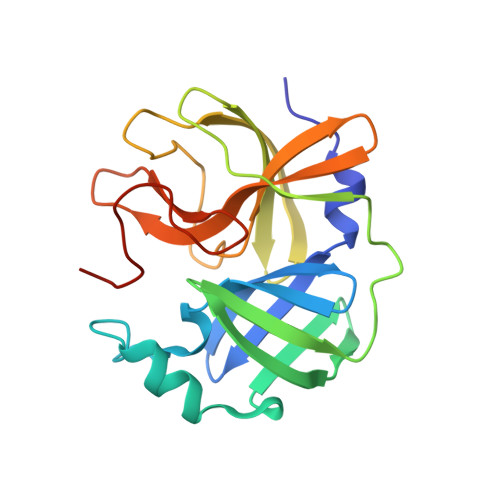
    The monomeric catalytic domain (residues 1-199) of SARS-CoV-2 main protease (MPro 1-199 ) fused to 25 amino acids of its flanking nsp4 region mediates its autoprocessing at the nsp4-MPro 1-199 junction. We report the catalytic activity and the dissociation constants of MPro 1-199 and its analogs with the covalent inhibitors GC373 and nirmatrelvir (NMV), and the estimated monomer-dimer equilibrium constants of these complexes. Mass spectrometry indicates the presence of the accumulated adduct of NMV bound to MPro WT and MPro 1-199 and not of GC373. A room temperature crystal structure reveals a native-like fold of the catalytic domain with an unwound oxyanion loop (E state). In contrast, the structure of a covalent complex of the catalytic domain-GC373 or NMV shows an oxyanion loop conformation (E* state) resembling the full-length mature dimer. These results suggest that the E-E* equilibrium modulates autoprocessing of the main protease when converting from a monomeric polyprotein precursor to the mature dimer.


  • Organizational Affiliation

    Laboratory of Chemical Physics, National Institute of Diabetes and Digestive and Kidney Diseases, National Institutes of Health, DHHS, Bethesda, MD, 20892-0520, USA.


Macromolecules
Find similar proteins by:  (by identity cutoff)  |  3D Structure
Entity ID: 1
MoleculeChains Sequence LengthOrganismDetailsImage
3C-like proteinase nsp5
A, B
191Severe acute respiratory syndrome coronavirus 2Mutation(s): 0 
EC: 3.4.22.69
UniProt
Find proteins for P0DTD1 (Severe acute respiratory syndrome coronavirus 2)
Explore P0DTD1 
Go to UniProtKB:  P0DTD1
Entity Groups  
Sequence Clusters30% Identity50% Identity70% Identity90% Identity95% Identity100% Identity
UniProt GroupP0DTD1
Sequence Annotations
Expand
  • Reference Sequence
Small Molecules
Ligands 1 Unique
IDChains Name / Formula / InChI Key2D Diagram3D Interactions
K36 (Subject of Investigation/LOI)
Query on K36

Download Ideal Coordinates CCD File 
C [auth A],
D [auth B]
(1S,2S)-2-({N-[(benzyloxy)carbonyl]-L-leucyl}amino)-1-hydroxy-3-[(3S)-2-oxopyrrolidin-3-yl]propane-1-sulfonic acid
C21 H31 N3 O8 S
BSPZFJDYQHDZNR-HTCLRFROSA-N
Biologically Interesting Molecules (External Reference) 1 Unique
Experimental Data & Validation

Experimental Data

  • Method: X-RAY DIFFRACTION
  • Resolution: 1.80 Å
  • R-Value Free: 0.179 
  • R-Value Work: 0.142 
  • R-Value Observed: 0.144 
  • Space Group: P 1 21 1
Unit Cell:
Length ( Å )Angle ( ˚ )
a = 52.792α = 90
b = 61.933β = 97.84
c = 58.939γ = 90
Software Package:
Software NamePurpose
PHENIXrefinement
CrysalisProdata reduction
CrysalisProdata scaling
PHASERphasing

Structure Validation

View Full Validation Report


Ligand Structure Quality Assessment 


Entry History & Funding Information

Deposition Data


Funding OrganizationLocationGrant Number
Not funded--

Revision History  (Full details and data files)

  • Version 1.0: 2022-10-05
    Type: Initial release
  • Version 1.1: 2023-10-18
    Changes: Data collection, Refinement description