7UIS | pdb_00007uis

Cocrystal structure of human CaMKII-alpha (CAMK2A)kinase domain and GluN2B(S1303D)


Experimental Data Snapshot

  • Method: X-RAY DIFFRACTION
  • Resolution: 2.58 Å
  • R-Value Free: 
    0.268 (Depositor), 0.270 (DCC) 
  • R-Value Work: 
    0.217 (Depositor), 0.222 (DCC) 
  • R-Value Observed: 
    0.220 (Depositor) 

Starting Model: experimental
View more details

wwPDB Validation 3D Report Full Report

Validation slider image for 7UIS

This is version 1.2 of the entry. See complete history

Literature

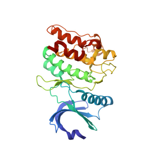
CaMKII binds both substrates and activators at the active site.

Ozden, C.Sloutsky, R.Mitsugi, T.Santos, N.Agnello, E.Gaubitz, C.Foster, J.Lapinskas, E.Esposito, E.A.Saneyoshi, T.Kelch, B.A.Garman, S.C.Hayashi, Y.Stratton, M.M.

(2022) Cell Rep 40: 111064-111064

  • DOI: https://doi.org/10.1016/j.celrep.2022.111064
  • Primary Citation Related Structures: 
    6X5G, 6X5Q, 7KL0, 7KL1, 7UIQ, 7UIR, 7UIS, 7UJP, 7UJQ, 7UJR, 7UJS, 7UJT

  • PubMed Abstract: 

    Ca 2+ /calmodulin-dependent protein kinase II (CaMKII) is a signaling protein required for long-term memory. When activated by Ca 2+ /CaM, it sustains activity even after the Ca 2+ dissipates. In addition to the well-known autophosphorylation-mediated mechanism, interaction with specific binding partners also persistently activates CaMKII. A long-standing model invokes two distinct S and T sites. If an interactor binds at the T-site, then it will preclude autoinhibition and allow substrates to be phosphorylated at the S site. Here, we specifically test this model with X-ray crystallography, molecular dynamics simulations, and biochemistry. Our data are inconsistent with this model. Co-crystal structures of four different activators or substrates show that they all bind to a single continuous site across the kinase domain. We propose a mechanistic model where persistent CaMKII activity is facilitated by high-affinity binding partners that kinetically compete with autoinhibition by the regulatory segment to allow substrate phosphorylation.


  • Organizational Affiliation
    • Department of Biochemistry and Molecular Biology, University of Massachusetts, Amherst, MA 01003, USA; Molecular and Cellular Biology Graduate Program, University of Massachusetts, Amherst, MA 01003, USA.

Macromolecule Content 

  • Total Structure Weight: 33.33 kDa 
  • Atom Count: 2,207 
  • Modeled Residue Count: 279 
  • Deposited Residue Count: 290 
  • Unique protein chains: 2

Macromolecules

Find similar proteins by:|  3D Structure
Entity ID: 1
MoleculeChains  Sequence LengthOrganismDetailsImage
Calcium/calmodulin-dependent protein kinase type II subunit alpha268Homo sapiensMutation(s): 2 
Gene Names: CAMK2ACAMKAKIAA0968
EC: 2.7.11.17
UniProt & NIH Common Fund Data Resources
Find proteins for Q9UQM7 (Homo sapiens)
Explore Q9UQM7 
Go to UniProtKB:  Q9UQM7
PHAROS:  Q9UQM7
GTEx:  ENSG00000070808 
Entity Groups
Sequence Clusters30% Identity50% Identity70% Identity90% Identity95% Identity100% Identity
UniProt GroupQ9UQM7
Sequence Annotations
Expand
Reference Sequence
Find similar proteins by:  Sequence   |   3D Structure  
Entity ID: 2
MoleculeChains  Sequence LengthOrganismDetailsImage
Glutamate receptor ionotropic, NMDA 2B22Homo sapiensMutation(s): 1 
UniProt & NIH Common Fund Data Resources
Find proteins for Q13224 (Homo sapiens)
Explore Q13224 
Go to UniProtKB:  Q13224
PHAROS:  Q13224
GTEx:  ENSG00000273079 
Entity Groups
Sequence Clusters30% Identity50% Identity70% Identity90% Identity95% Identity100% Identity
UniProt GroupQ13224
Sequence Annotations
Expand
Reference Sequence

Experimental Data & Validation

Experimental Data

  • Method: X-RAY DIFFRACTION
  • Resolution: 2.58 Å
  • R-Value Free:  0.268 (Depositor), 0.270 (DCC) 
  • R-Value Work:  0.217 (Depositor), 0.222 (DCC) 
  • R-Value Observed: 0.220 (Depositor) 
Space Group: P 1 21 1
Unit Cell:
Length ( Å )Angle ( ˚ )
a = 43.413α = 90
b = 71.426β = 97.51
c = 45.276γ = 90
Software Package:
Software NamePurpose
REFMACrefinement
HKL-2000data reduction
HKL-2000data scaling
PHASERphasing
PDB_EXTRACTdata extraction

Structure Validation

View Full Validation Report


Entry History 

& Funding Information

Deposition Data


Funding OrganizationLocationGrant Number
Not funded--

Revision History  (Full details and data files)

  • Version 1.0: 2022-04-06
    Type: Initial release
  • Version 1.1: 2022-08-10
    Changes: Database references
  • Version 1.2: 2023-10-18
    Changes: Data collection, Refinement description