7UGG | pdb_00007ugg

Cryo-EM structure of TRPV3 in complex with the anesthetic dyclonine

  • Classification: MEMBRANE PROTEIN
  • Organism(s): Mus musculus
  • Expression System: Homo sapiens
  • Mutation(s): No 
  • Membrane Protein: Yes  OPMPDBTMmpstruc

  • Deposited: 2022-03-24 Released: 2022-06-08 
  • Deposition Author(s): Neuberger, A., Nadezhdin, K.D., Sobolevsky, A.I.
  • Funding Organization(s): National Institutes of Health/National Cancer Institute (NIH/NCI), National Institutes of Health/National Institute of Neurological Disorders and Stroke (NIH/NINDS), National Science Foundation (NSF, United States), German Research Foundation (DFG)

Experimental Data Snapshot

  • Method: ELECTRON MICROSCOPY
  • Resolution: 3.16 Å
  • Aggregation State: PARTICLE 
  • Reconstruction Method: SINGLE PARTICLE 

Starting Model: experimental
View more details

wwPDB Validation 3D Report Full Report

Validation slider image for 7UGG

This is version 1.1 of the entry. See complete history

Literature

Structural mechanism of TRPV3 channel inhibition by the anesthetic dyclonine.

Neuberger, A.Nadezhdin, K.D.Sobolevsky, A.I.

(2022) Nat Commun 13: 2795-2795

  • DOI: https://doi.org/10.1038/s41467-022-30537-8
  • Primary Citation Related Structures: 
    7UGG

  • PubMed Abstract: 

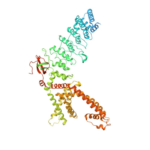
    Skin diseases are common human illnesses that occur in all cultures, at all ages, and affect between 30% and 70% of individuals globally. TRPV3 is a cation-permeable TRP channel predominantly expressed in skin keratinocytes, implicated in cutaneous sensation and associated with numerous skin diseases. TRPV3 is inhibited by the local anesthetic dyclonine, traditionally used for topical applications to relieve pain and itch. However, the structural basis of TRPV3 inhibition by dyclonine has remained elusive. Here we present a cryo-EM structure of a TRPV3-dyclonine complex that reveals binding of the inhibitor in the portals which connect the membrane environment surrounding the channel to the central cavity of the channel pore. We propose a mechanism of TRPV3 inhibition in which dyclonine molecules stick out into the channel pore, creating a barrier for ion conductance. The allosteric binding site of dyclonine can serve as a template for the design of new TRPV3-targeting drugs.


  • Organizational Affiliation
    • Department of Biochemistry and Molecular Biophysics, Columbia University, New York, NY, USA.

Macromolecule Content 

  • Total Structure Weight: 389.74 kDa 
  • Atom Count: 21,670 
  • Modeled Residue Count: 2,480 
  • Deposited Residue Count: 3,232 
  • Unique protein chains: 1

Macromolecules

Find similar proteins by:|  3D Structure
Entity ID: 1
MoleculeChains  Sequence LengthOrganismDetailsImage
Transient receptor potential cation channel subfamily V member 3
A, B, C, D
808Mus musculusMutation(s): 0 
Gene Names: Trpv3
Membrane Entity: Yes 
UniProt
Find proteins for Q8K424 (Mus musculus)
Explore Q8K424 
Go to UniProtKB:  Q8K424
Entity Groups
Sequence Clusters30% Identity50% Identity70% Identity90% Identity95% Identity100% Identity
UniProt GroupQ8K424
Sequence Annotations
Expand
Reference Sequence

Small Molecules

Ligands 3 Unique
IDChains Name / Formula / InChI Key2D Diagram3D Interactions
POV

Query on POV


Download:Ideal Coordinates CCD File
AA [auth D]
BA [auth D]
CA [auth D]
DA [auth D]
E [auth A]
AA [auth D],
BA [auth D],
CA [auth D],
DA [auth D],
E [auth A],
EA [auth D],
F [auth A],
FA [auth D],
G [auth A],
H [auth A],
J [auth A],
L [auth A],
N [auth B],
O [auth B],
P [auth B],
Q [auth B],
R [auth B],
S [auth B],
U [auth C],
V [auth C],
W [auth C],
X [auth C],
Y [auth C],
Z [auth C]
(2S)-3-(hexadecanoyloxy)-2-[(9Z)-octadec-9-enoyloxy]propyl 2-(trimethylammonio)ethyl phosphate
C42 H82 N O8 P
WTJKGGKOPKCXLL-PFDVCBLKSA-N
N8R
(Subject of Investigation/LOI)

Query on N8R


Download:Ideal Coordinates CCD File
GA [auth D],
I [auth A],
K [auth A],
T [auth B]
Dyclonine
C18 H27 N O2
BZEWSEKUUPWQDQ-UHFFFAOYSA-N
NA

Query on NA


Download:Ideal Coordinates CCD File
HA [auth D],
M [auth A]
SODIUM ION
Na
FKNQFGJONOIPTF-UHFFFAOYSA-N
Binding Affinity Annotations 
IDSourceBinding Affinity
N8R BindingDB:  7UGG IC50: 3200 (nM) from 1 assay(s)

Experimental Data & Validation

Experimental Data

  • Method: ELECTRON MICROSCOPY
  • Resolution: 3.16 Å
  • Aggregation State: PARTICLE 
  • Reconstruction Method: SINGLE PARTICLE 
EM Software:
TaskSoftware PackageVersion
RECONSTRUCTIONcryoSPARC3.1
MODEL REFINEMENTPHENIX

Structure Validation

View Full Validation Report


Entry History 

& Funding Information

Deposition Data


Funding OrganizationLocationGrant Number
National Institutes of Health/National Cancer Institute (NIH/NCI)United StatesR01 CA206573
National Institutes of Health/National Institute of Neurological Disorders and Stroke (NIH/NINDS)United StatesR01 NS083660
National Institutes of Health/National Institute of Neurological Disorders and Stroke (NIH/NINDS)United StatesR01 NS107253
National Science Foundation (NSF, United States)United States1818086
German Research Foundation (DFG)Germany464295817

Revision History  (Full details and data files)

  • Version 1.0: 2022-06-08
    Type: Initial release
  • Version 1.1: 2024-02-14
    Changes: Data collection, Refinement description