7UGB | pdb_00007ugb

Crystal structure of rat ERK2 complexed with docking peptide from ISG20


Experimental Data Snapshot

  • Method: X-RAY DIFFRACTION
  • Resolution: 1.90 Å
  • R-Value Free: 
    0.228 (Depositor), 0.230 (DCC) 
  • R-Value Work: 
    0.179 (Depositor), 0.180 (DCC) 
  • R-Value Observed: 
    0.181 (Depositor) 

Starting Model: experimental
View more details

wwPDB Validation   3D Report Full Report


Ligand Structure Quality Assessment 


This is version 1.3 of the entry. See complete history


Literature

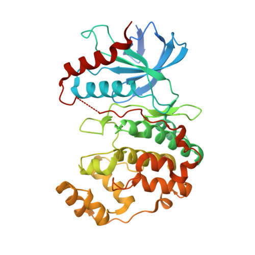
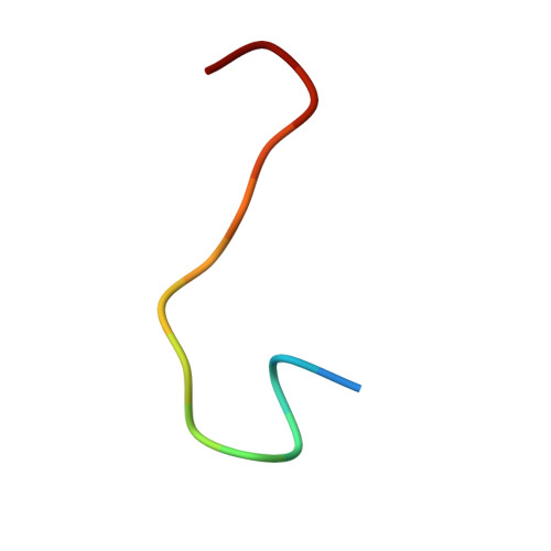
Cancer hotspot mutations rewire ERK2 specificity by selective exclusion of docking interactions.

Torres Robles, J.Stiegler, A.L.Boggon, T.J.Turk, B.E.

(2025) J Biological Chem 301: 108348-108348

  • DOI: https://doi.org/10.1016/j.jbc.2025.108348
  • Primary Citation of Related Structures:  
    7UGB

  • PubMed Abstract: 

    The protein kinase ERK2 is recurrently mutated in human squamous cell carcinomas and other tumors. ERK2 mutations cluster in an essential docking recruitment site that interacts with short linear motifs found within intrinsically disordered regions of ERK substrates and regulators. Cancer-associated mutations do not disrupt ERK2 docking interactions altogether but selectively inhibit some interactions while sparing others. However, the full scope of disrupted or maintained interactions remains unknown, limiting our understanding of how these mutations contribute to cancer. We recently defined the docking interactome of wild-type ERK2 by screening a yeast two-hybrid library of proteomic short linear motifs. Here, we apply this approach to the two most recurrent cancer-associated mutants. We find that most sequences binding to WT ERK2 also interact with both mutant forms. Analysis of differentially interacting sequences revealed that ERK2 mutants selectively lose the ability to bind sequences conforming to a specific motif. We solved the co-crystal structure of ERK2 in complex with a peptide fragment of ISG20, a screening hit that binds exclusively to the WT kinase. This structure demonstrated the mechanism by which cancer hotspot mutations at Glu81, Arg135, Asp321, and Glu322 selectively impact peptide binding. Finally, we found that cancer-associated ERK2 mutations had decreased activity in phosphorylating GEF-H1/ARHGEF2, a known ERK substrate harboring a WT-selective docking motif. Collectively, our studies provide a structural rationale for how a broad set of interactions are disrupted by ERK2 hotspot mutations, suggesting mechanisms for pathway rewiring in cancers harboring these mutations.


  • Organizational Affiliation
    • Department of Chemistry, Yale University, New Haven, Connecticut, USA; Department of Pharmacology, Yale School of Medicine, New Haven, Connecticut, USA.

Macromolecules
Find similar proteins by:  (by identity cutoff)  |  3D Structure
Entity ID: 1
MoleculeChains Sequence LengthOrganismDetailsImage
Mitogen-activated protein kinase 1380Rattus norvegicusMutation(s): 0 
Gene Names: Mapk1Erk2MapkPrkm1
EC: 2.7.11.24
UniProt
Find proteins for P63086 (Rattus norvegicus)
Explore P63086 
Go to UniProtKB:  P63086
Entity Groups  
Sequence Clusters30% Identity50% Identity70% Identity90% Identity95% Identity100% Identity
UniProt GroupP63086
Sequence Annotations
Expand
  • Reference Sequence

Find similar proteins by:  Sequence   |   3D Structure  

Entity ID: 2
MoleculeChains Sequence LengthOrganismDetailsImage
Interferon-stimulated gene 20 kDa proteinB [auth I]15Homo sapiensMutation(s): 1 
EC: 3.1.13.1
UniProt & NIH Common Fund Data Resources
Find proteins for Q96AZ6 (Homo sapiens)
Explore Q96AZ6 
Go to UniProtKB:  Q96AZ6
PHAROS:  Q96AZ6
GTEx:  ENSG00000172183 
Entity Groups  
Sequence Clusters30% Identity50% Identity70% Identity90% Identity95% Identity100% Identity
UniProt GroupQ96AZ6
Sequence Annotations
Expand
  • Reference Sequence
Small Molecules
Ligands 1 Unique
IDChains Name / Formula / InChI Key2D Diagram3D Interactions
ANP
Query on ANP

Download Ideal Coordinates CCD File 
C [auth A]PHOSPHOAMINOPHOSPHONIC ACID-ADENYLATE ESTER
C10 H17 N6 O12 P3
PVKSNHVPLWYQGJ-KQYNXXCUSA-N
Experimental Data & Validation

Experimental Data

  • Method: X-RAY DIFFRACTION
  • Resolution: 1.90 Å
  • R-Value Free:  0.228 (Depositor), 0.230 (DCC) 
  • R-Value Work:  0.179 (Depositor), 0.180 (DCC) 
  • R-Value Observed: 0.181 (Depositor) 
Space Group: P 21 21 21
Unit Cell:
Length ( Å )Angle ( ˚ )
a = 45.888α = 90
b = 66.679β = 90
c = 116.969γ = 90
Software Package:
Software NamePurpose
HKL-2000data scaling
PHASERphasing
PHENIXrefinement
PDB_EXTRACTdata extraction
HKL-2000data reduction

Structure Validation

View Full Validation Report


Ligand Structure Quality Assessment 


Entry History & Funding Information

Deposition Data


Funding OrganizationLocationGrant Number
National Institutes of Health/National Institute of General Medical Sciences (NIH/NIGMS)United StatesR01 GM135331
National Science Foundation (NSF, United States)United States1752134

Revision History  (Full details and data files)

  • Version 1.0: 2023-03-29
    Type: Initial release
  • Version 1.1: 2023-10-25
    Changes: Data collection, Refinement description
  • Version 1.2: 2024-10-23
    Changes: Structure summary
  • Version 1.3: 2025-04-02
    Changes: Database references