7UFZ

Crystal structure of TDP1 complexed with compound XZ768


Experimental Data Snapshot

  • Method: X-RAY DIFFRACTION
  • Resolution: 1.56 Å
  • R-Value Free: 0.195 
  • R-Value Work: 0.171 
  • R-Value Observed: 0.172 

wwPDB Validation   3D Report Full Report


Ligand Structure Quality Assessment 


This is version 1.1 of the entry. See complete history


Literature

Phosphonic acid-containing inhibitors of tyrosyl-DNA phosphodiesterase 1.

Zhao, X.Z.Wang, W.Lountos, G.T.Tropea, J.E.Needle, D.Pommier, Y.Burke Jr., T.R.

(2022) Front Chem 10: 910953-910953

  • DOI: https://doi.org/10.3389/fchem.2022.910953
  • Primary Citation of Related Structures:  
    7UFY, 7UFZ

  • PubMed Abstract: 

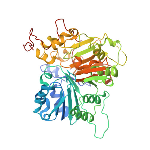
    Tyrosyl-DNA phosphodiesterase 1 (TDP1) repairs stalled type I topoisomerase (TOP1)-DNA complexes by hydrolyzing the phosphodiester bond between the TOP1 Y723 residue and the 3'-phosphate of its DNA substrate. Although TDP1 antagonists could potentially reduce the dose of TOP1 inhibitors needed to achieve effective anticancer effects, the development of validated TDP1 inhibitors has proven to be challenging. This may, in part, be due to the open and extended nature of the TOP1 substrate binding region. We have previously reported imidazopyrazines and imidazopyridines that can inhibit TDP1 catalytic function in vitro . We solved the TDP1 crystal structures with bound inhibitors of this class and found that the dicarboxylic acid functionality within the N -(3,4-dicarboxyphenyl)-2-diphenylimidazo [ 1,2-a ]pyridin-3-amine platform overlaps with aspects of phosphoryl substrate recognition. Yet phosphonic acids could potentially better-replicate cognate TOP1-DNA substrate binding interactions than carboxylic acids. As reported herein, we designed phosphonic acid-containing variants of our previously reported carboxylic acid-containing imidazopyrazine and imidazopyridine inhibitors and effected their synthesis using one-pot Groebke-Blackburn-Bienayme multicomponent reactions. We obtained crystal structures of TDP1 complexed with a subset of inhibitors. We discuss binding interactions of these inhibitors within the context of phosphate-containing substrate and carboxylic acid-based inhibitors. These compounds represent a new structural class of small molecule ligands that mimic aspects of the 3'-processed substrate that results from TDP1 catalysis.


  • Organizational Affiliation

    Chemical Biology Laboratory, Center for Cancer Research, National Cancer Institute, National Institutes of Health, Frederick, MD, United States.


Macromolecules
Find similar proteins by:  (by identity cutoff)  |  3D Structure
Entity ID: 1
MoleculeChains Sequence LengthOrganismDetailsImage
Tyrosyl-DNA phosphodiesterase 1
A, B
461Homo sapiensMutation(s): 0 
Gene Names: TDP1
EC: 3.1.4
UniProt & NIH Common Fund Data Resources
Find proteins for Q9NUW8 (Homo sapiens)
Explore Q9NUW8 
Go to UniProtKB:  Q9NUW8
PHAROS:  Q9NUW8
GTEx:  ENSG00000042088 
Entity Groups  
Sequence Clusters30% Identity50% Identity70% Identity90% Identity95% Identity100% Identity
UniProt GroupQ9NUW8
Sequence Annotations
Expand
  • Reference Sequence
Small Molecules
Ligands 3 Unique
IDChains Name / Formula / InChI Key2D Diagram3D Interactions
N7U (Subject of Investigation/LOI)
Query on N7U

Download Ideal Coordinates CCD File 
C [auth A],
D [auth A],
J [auth B],
K [auth B]
(4-{[(4S)-2-phenylimidazo[1,2-a]pyridin-3-yl]amino}phenyl)phosphonic acid
C19 H16 N3 O3 P
PVOVQXFFCUTLSZ-UHFFFAOYSA-N
DMS
Query on DMS

Download Ideal Coordinates CCD File 
I [auth A]DIMETHYL SULFOXIDE
C2 H6 O S
IAZDPXIOMUYVGZ-UHFFFAOYSA-N
EDO
Query on EDO

Download Ideal Coordinates CCD File 
E [auth A]
F [auth A]
G [auth A]
H [auth A]
L [auth B]
E [auth A],
F [auth A],
G [auth A],
H [auth A],
L [auth B],
M [auth B],
N [auth B]
1,2-ETHANEDIOL
C2 H6 O2
LYCAIKOWRPUZTN-UHFFFAOYSA-N
Experimental Data & Validation

Experimental Data

  • Method: X-RAY DIFFRACTION
  • Resolution: 1.56 Å
  • R-Value Free: 0.195 
  • R-Value Work: 0.171 
  • R-Value Observed: 0.172 
  • Space Group: P 21 21 21
Unit Cell:
Length ( Å )Angle ( ˚ )
a = 49.889α = 90
b = 104.604β = 90
c = 193.095γ = 90
Software Package:
Software NamePurpose
PHENIXrefinement
PDB_EXTRACTdata extraction
HKL-3000data reduction
HKL-3000data scaling
PHASERphasing

Structure Validation

View Full Validation Report


Ligand Structure Quality Assessment 


Entry History & Funding Information

Deposition Data


Funding OrganizationLocationGrant Number
National Institutes of Health/National Cancer Institute (NIH/NCI)United States--

Revision History  (Full details and data files)

  • Version 1.0: 2023-04-12
    Type: Initial release
  • Version 1.1: 2023-10-25
    Changes: Data collection, Refinement description