De novo design of modular peptide-binding proteins by superhelical matching.
Wu, K., Bai, H., Chang, Y.T., Redler, R., McNally, K.E., Sheffler, W., Brunette, T.J., Hicks, D.R., Morgan, T.E., Stevens, T.J., Broerman, A., Goreshnik, I., DeWitt, M., Chow, C.M., Shen, Y., Stewart, L., Derivery, E., Silva, D.A., Bhabha, G., Ekiert, D.C., Baker, D.(2023) Nature 616: 581-589
- PubMed: 37020023 Search on PubMedSearch on PubMed Central
- DOI: https://doi.org/10.1038/s41586-023-05909-9
- Primary Citation Related Structures:
7UDJ, 7UDK, 7UDL, 7UDM, 7UDN, 7UDO, 7UE2 - PubMed Abstract:
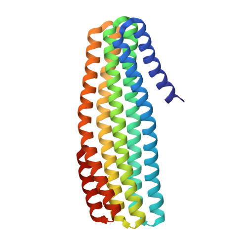
General approaches for designing sequence-specific peptide-binding proteins would have wide utility in proteomics and synthetic biology. However, designing peptide-binding proteins is challenging, as most peptides do not have defined structures in isolation, and hydrogen bonds must be made to the buried polar groups in the peptide backbone 1-3 . Here, inspired by natural and re-engineered protein-peptide systems 4-11 , we set out to design proteins made out of repeating units that bind peptides with repeating sequences, with a one-to-one correspondence between the repeat units of the protein and those of the peptide. We use geometric hashing to identify protein backbones and peptide-docking arrangements that are compatible with bidentate hydrogen bonds between the side chains of the protein and the peptide backbone 12 . The remainder of the protein sequence is then optimized for folding and peptide binding. We design repeat proteins to bind to six different tripeptide-repeat sequences in polyproline II conformations. The proteins are hyperstable and bind to four to six tandem repeats of their tripeptide targets with nanomolar to picomolar affinities in vitro and in living cells. Crystal structures reveal repeating interactions between protein and peptide interactions as designed, including ladders of hydrogen bonds from protein side chains to peptide backbones. By redesigning the binding interfaces of individual repeat units, specificity can be achieved for non-repeating peptide sequences and for disordered regions of native proteins.
- Department of Biochemistry, University of Washington, Seattle, WA, USA.
Organizational Affiliation: