7UD7

Crystal structure of deoxygenated hemoglobin in complex with 5HMF-NO at 1.8 Angstrom

  • Classification: OXYGEN TRANSPORT
  • Organism(s): Homo sapiens
  • Expression System: Homo sapiens
  • Mutation(s): No 

  • Deposited: 2022-03-18 Released: 2022-03-30 
  • Deposition Author(s): Donkor, A.K., Musayev, F.N., Safo, M.K.
  • Funding Organization(s): National Institutes of Health/National Institute on Minority Health and Health Disparities (NIH/NIMHD), National Institutes of Health/National Heart, Lung, and Blood Institute (NIH/NHLBI), National Institutes of Health/National Center for Research Resources (NIH/NCRR)

Experimental Data Snapshot

  • Method: X-RAY DIFFRACTION
  • Resolution: 1.80 Å
  • R-Value Free: 0.191 
  • R-Value Work: 0.151 
  • R-Value Observed: 0.153 

wwPDB Validation   3D Report Full Report


Ligand Structure Quality Assessment 


This is version 1.3 of the entry. See complete history


Literature

Design, Synthesis, and Antisickling Investigation of a Nitric Oxide-Releasing Prodrug of 5HMF for the Treatment of Sickle Cell Disease.

Alhashimi, R.T.Ghatge, M.S.Donkor, A.K.Deshpande, T.M.Anabaraonye, N.Alramadhani, D.Danso-Danquah, R.Huang, B.Zhang, Y.Musayev, F.N.Abdulmalik, O.Safo, M.K.

(2022) Biomolecules 12

  • DOI: https://doi.org/10.3390/biom12050696
  • Primary Citation of Related Structures:  
    7UD7, 7UD8

  • PubMed Abstract: 

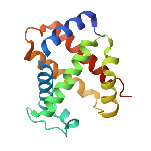
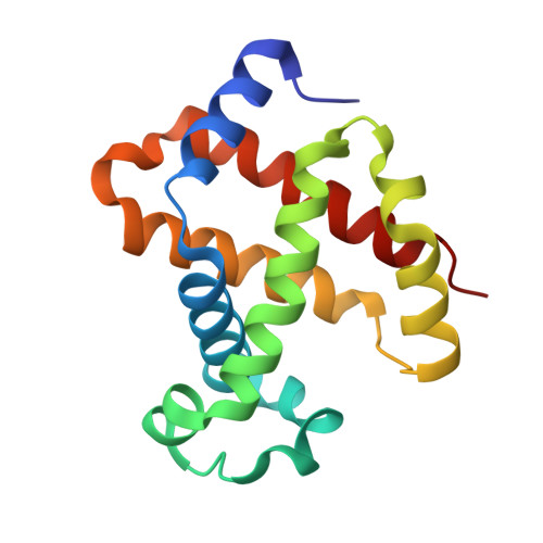
    5-hydroxyfurfural (5HMF), an allosteric effector of hemoglobin (Hb) with an ability to increase Hb affinity for oxygen has been studied extensively for its antisickling effect in vitro and in vivo, and in humans for the treatment of sickle cell disease (SCD). One of the downstream pathophysiologies of SCD is nitric oxide (NO) deficiency, therefore increasing NO (bio)availability is known to mitigate the severity of SCD symptoms. We report the synthesis of an NO-releasing prodrug of 5HMF (5HMF-NO), which in vivo, is expected to be bio-transformed into 5HMF and NO, with concomitant therapeutic activities. In vitro studies showed that when incubated with whole blood, 5HMF-NO releases NO, as anticipated. When incubated with sickle blood, 5HMF-NO formed Schiff base adduct with Hb, increased Hb affinity for oxygen, and prevented hypoxia-induced erythrocyte sickling, which at 1 mM concentration were 16%, 10% and 27%, respectively, compared to 21%, 18% and 21% for 5HMF. Crystal structures of 5HMF-NO with Hb showed 5HMF-NO bound to unliganded (deoxygenated) Hb, while the hydrolyzed product, 5HMF bound to liganded (carbonmonoxy-ligated) Hb. Our findings from this proof-of-concept study suggest that the incorporation of NO donor group to 5HMF and analogous molecules could be a novel beneficial strategy to treat SCD and warrants further detailed in vivo studies.


  • Organizational Affiliation

    Department of Medicinal Chemistry and The Institute for Structural Biology, Drug Discovery and Development, School of Pharmacy, Virginia Commonwealth University, Richmond, VA 23298, USA.


Macromolecules
Find similar proteins by:  (by identity cutoff)  |  3D Structure
Entity ID: 1
MoleculeChains Sequence LengthOrganismDetailsImage
Hemoglobin subunit alpha
A, C
142Homo sapiensMutation(s): 0 
Gene Names: HBA1HBA2
UniProt & NIH Common Fund Data Resources
Find proteins for P69905 (Homo sapiens)
Explore P69905 
Go to UniProtKB:  P69905
PHAROS:  P69905
Entity Groups  
Sequence Clusters30% Identity50% Identity70% Identity90% Identity95% Identity100% Identity
UniProt GroupP69905
Sequence Annotations
Expand
  • Reference Sequence
Find similar proteins by:  (by identity cutoff)  |  3D Structure
Entity ID: 2
MoleculeChains Sequence LengthOrganismDetailsImage
Hemoglobin subunit beta
B, D
147Homo sapiensMutation(s): 0 
Gene Names: HBB
UniProt & NIH Common Fund Data Resources
Find proteins for P68871 (Homo sapiens)
Explore P68871 
Go to UniProtKB:  P68871
PHAROS:  P68871
GTEx:  ENSG00000244734 
Entity Groups  
Sequence Clusters30% Identity50% Identity70% Identity90% Identity95% Identity100% Identity
UniProt GroupP68871
Sequence Annotations
Expand
  • Reference Sequence
Experimental Data & Validation

Experimental Data

  • Method: X-RAY DIFFRACTION
  • Resolution: 1.80 Å
  • R-Value Free: 0.191 
  • R-Value Work: 0.151 
  • R-Value Observed: 0.153 
  • Space Group: P 1 21 1
Unit Cell:
Length ( Å )Angle ( ˚ )
a = 62.344α = 90
b = 82.031β = 99.49
c = 53.454γ = 90
Software Package:
Software NamePurpose
d*TREKdata scaling
PHENIXrefinement
PDB_EXTRACTdata extraction
CrysalisProdata reduction
PHASERphasing

Structure Validation

View Full Validation Report


Ligand Structure Quality Assessment 


Entry History & Funding Information

Deposition Data


Funding OrganizationLocationGrant Number
National Institutes of Health/National Institute on Minority Health and Health Disparities (NIH/NIMHD)United StatesR01MD009124
National Institutes of Health/National Heart, Lung, and Blood Institute (NIH/NHLBI)United StatesR61HL156158
National Institutes of Health/National Center for Research Resources (NIH/NCRR)United StatesS10OD021756

Revision History  (Full details and data files)

  • Version 1.0: 2022-03-30
    Type: Initial release
  • Version 1.1: 2022-04-06
    Changes: Structure summary
  • Version 1.2: 2022-06-29
    Changes: Database references
  • Version 1.3: 2023-10-18
    Changes: Data collection, Refinement description