7UD5

Complex between MLL1-WRAD and an H2B-ubiquitinated nucleosome


Experimental Data Snapshot

  • Method: ELECTRON MICROSCOPY
  • Resolution: 4.25 Å
  • Aggregation State: PARTICLE 
  • Reconstruction Method: SINGLE PARTICLE 

wwPDB Validation   3D Report Full Report


This is version 1.2 of the entry. See complete history

Re-refinement Note

This entry reflects an alternative modeling of the original data in: 8DU4


Literature

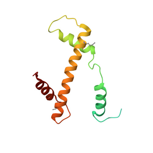
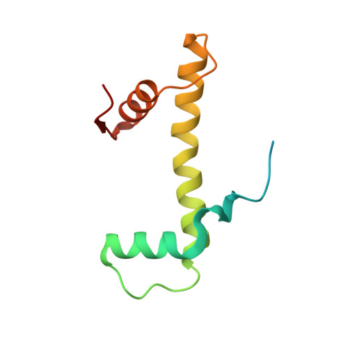
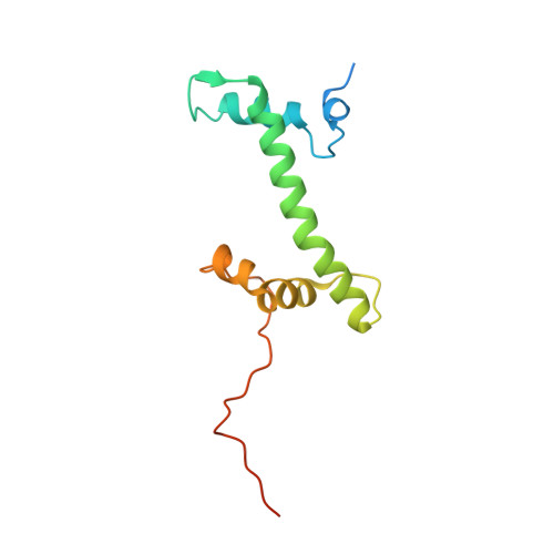
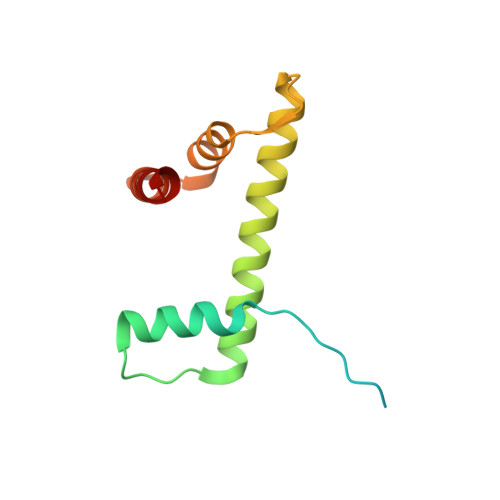
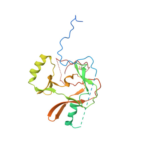
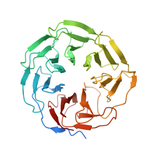
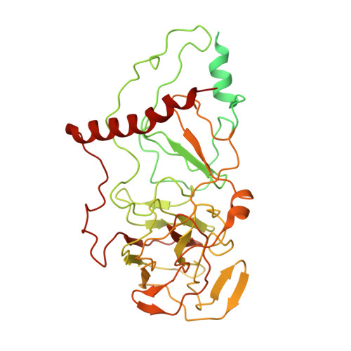
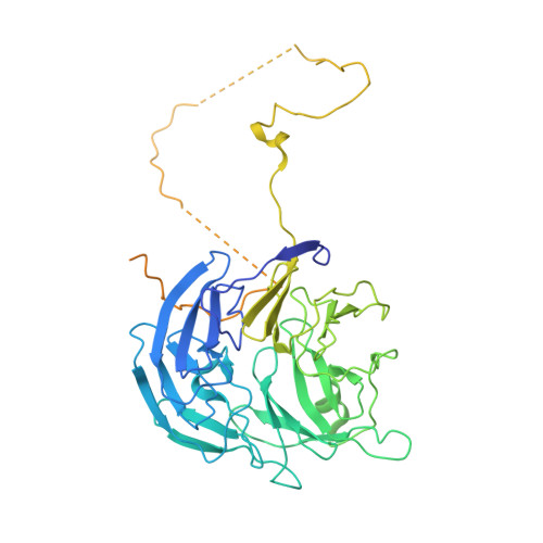
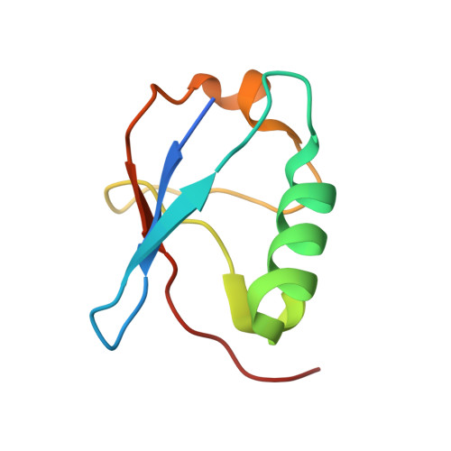
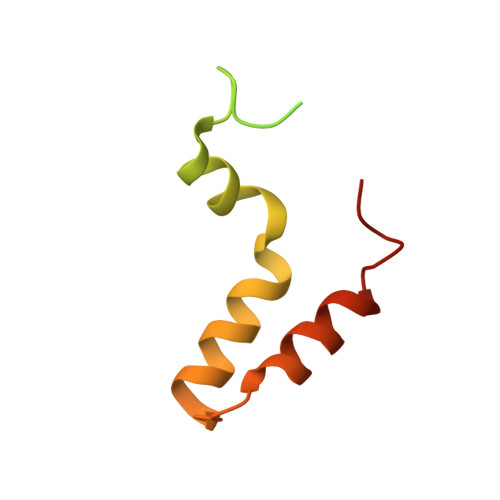
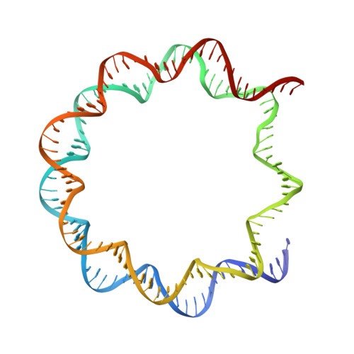
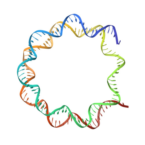
Multistate structures of the MLL1-WRAD complex bound to H2B-ubiquitinated nucleosome.

Rahman, S.Hoffmann, N.A.Worden, E.J.Smith, M.L.Namitz, K.E.W.Knutson, B.A.Cosgrove, M.S.Wolberger, C.

(2022) Proc Natl Acad Sci U S A 119: e2205691119-e2205691119

  • DOI: https://doi.org/10.1073/pnas.2205691119
  • Primary Citation of Related Structures:  
    7UD5, 8DU4

  • PubMed Abstract: 

    The human Mixed Lineage Leukemia-1 (MLL1) complex methylates histone H3K4 to promote transcription and is stimulated by monoubiquitination of histone H2B. Recent structures of the MLL1-WRAD core complex, which comprises the MLL1 methyltransferase, W DR5, R bBp5, A sh2L, and D PY-30, have revealed variability in the docking of MLL1-WRAD on nucleosomes. In addition, portions of the Ash2L structure and the position of DPY30 remain ambiguous. We used an integrated approach combining cryoelectron microscopy (cryo-EM) and mass spectrometry cross-linking to determine a structure of the MLL1-WRAD complex bound to ubiquitinated nucleosomes. The resulting model contains the Ash2L intrinsically disordered region (IDR), SPRY insertion region, Sdc1-DPY30 interacting region (SDI-motif), and the DPY30 dimer. We also resolved three additional states of MLL1-WRAD lacking one or more subunits, which may reflect different steps in the assembly of MLL1-WRAD. The docking of subunits in all four states differs from structures of MLL1-WRAD bound to unmodified nucleosomes, suggesting that H2B-ubiquitin favors assembly of the active complex. Our results provide a more complete picture of MLL1-WRAD and the role of ubiquitin in promoting formation of the active methyltransferase complex.


  • Organizational Affiliation

    Department of Biophysics and Biophysical Chemistry, The Johns Hopkins University School of Medicine, Baltimore, MD 21205.


Macromolecules

Find similar proteins by:  (by identity cutoff)  |  3D Structure
Entity ID: 1
MoleculeChains Sequence LengthOrganismDetailsImage
Histone H3
A, E
136Xenopus laevisMutation(s): 3 
Gene Names: XELAEV_18002543mg
UniProt
Find proteins for P84233 (Xenopus laevis)
Explore P84233 
Go to UniProtKB:  P84233
Entity Groups  
Sequence Clusters30% Identity50% Identity70% Identity90% Identity95% Identity100% Identity
UniProt GroupP84233
Sequence Annotations
Expand
  • Reference Sequence
Find similar proteins by:  (by identity cutoff)  |  3D Structure
Entity ID: 2
MoleculeChains Sequence LengthOrganismDetailsImage
Histone H4
B, F
103Xenopus laevisMutation(s): 0 
UniProt
Find proteins for P62799 (Xenopus laevis)
Explore P62799 
Go to UniProtKB:  P62799
Entity Groups  
Sequence Clusters30% Identity50% Identity70% Identity90% Identity95% Identity100% Identity
UniProt GroupP62799
Sequence Annotations
Expand
  • Reference Sequence
Find similar proteins by:  (by identity cutoff)  |  3D Structure
Entity ID: 3
MoleculeChains Sequence LengthOrganismDetailsImage
Histone H2A
C, G
130Xenopus laevisMutation(s): 0 
Gene Names: hist1h2ajLOC494591XELAEV_18003602mg
UniProt
Find proteins for P06897 (Xenopus laevis)
Explore P06897 
Go to UniProtKB:  P06897
Entity Groups  
Sequence Clusters30% Identity50% Identity70% Identity90% Identity95% Identity100% Identity
UniProt GroupP06897
Sequence Annotations
Expand
  • Reference Sequence
Find similar proteins by:  (by identity cutoff)  |  3D Structure
Entity ID: 4
MoleculeChains Sequence LengthOrganismDetailsImage
Histone H2B 1.1
D, H
123Xenopus laevisMutation(s): 2 
UniProt
Find proteins for P02281 (Xenopus laevis)
Explore P02281 
Go to UniProtKB:  P02281
Entity Groups  
Sequence Clusters30% Identity50% Identity70% Identity90% Identity95% Identity100% Identity
UniProt GroupP02281
Sequence Annotations
Expand
  • Reference Sequence
Find similar proteins by:  (by identity cutoff)  |  3D Structure
Entity ID: 7
MoleculeChains Sequence LengthOrganismDetailsImage
cDNA FLJ56846, highly similar to Zinc finger protein HRX226Homo sapiensMutation(s): 0 
UniProt
Find proteins for B4DIJ7 (Homo sapiens)
Explore B4DIJ7 
Go to UniProtKB:  B4DIJ7
Entity Groups  
Sequence Clusters30% Identity50% Identity70% Identity90% Identity95% Identity100% Identity
UniProt GroupB4DIJ7
Sequence Annotations
Expand
  • Reference Sequence
Find similar proteins by:  (by identity cutoff)  |  3D Structure
Entity ID: 8
MoleculeChains Sequence LengthOrganismDetailsImage
WD repeat-containing protein 5335Homo sapiensMutation(s): 0 
Gene Names: WDR5BIG3
UniProt & NIH Common Fund Data Resources
Find proteins for P61964 (Homo sapiens)
Explore P61964 
Go to UniProtKB:  P61964
PHAROS:  P61964
GTEx:  ENSG00000196363 
Entity Groups  
Sequence Clusters30% Identity50% Identity70% Identity90% Identity95% Identity100% Identity
UniProt GroupP61964
Sequence Annotations
Expand
  • Reference Sequence
Find similar proteins by:  (by identity cutoff)  |  3D Structure
Entity ID: 9
MoleculeChains Sequence LengthOrganismDetailsImage
Set1/Ash2 histone methyltransferase complex subunit ASH2534Homo sapiensMutation(s): 0 
Gene Names: ASH2LASH2L1
UniProt & NIH Common Fund Data Resources
Find proteins for Q9UBL3 (Homo sapiens)
Explore Q9UBL3 
Go to UniProtKB:  Q9UBL3
PHAROS:  Q9UBL3
GTEx:  ENSG00000129691 
Entity Groups  
Sequence Clusters30% Identity50% Identity70% Identity90% Identity95% Identity100% Identity
UniProt GroupQ9UBL3
Sequence Annotations
Expand
  • Reference Sequence
Find similar proteins by:  (by identity cutoff)  |  3D Structure
Entity ID: 10
MoleculeChains Sequence LengthOrganismDetailsImage
Retinoblastoma-binding protein 5538Homo sapiensMutation(s): 0 
Gene Names: RBBP5RBQ3
UniProt & NIH Common Fund Data Resources
Find proteins for Q15291 (Homo sapiens)
Explore Q15291 
Go to UniProtKB:  Q15291
PHAROS:  Q15291
GTEx:  ENSG00000117222 
Entity Groups  
Sequence Clusters30% Identity50% Identity70% Identity90% Identity95% Identity100% Identity
UniProt GroupQ15291
Sequence Annotations
Expand
  • Reference Sequence
Find similar proteins by:  (by identity cutoff)  |  3D Structure
Entity ID: 11
MoleculeChains Sequence LengthOrganismDetailsImage
Polyubiquitin-B81Homo sapiensMutation(s): 1 
Gene Names: UBB
UniProt & NIH Common Fund Data Resources
Find proteins for P0CG47 (Homo sapiens)
Explore P0CG47 
Go to UniProtKB:  P0CG47
PHAROS:  P0CG47
GTEx:  ENSG00000170315 
Entity Groups  
Sequence Clusters30% Identity50% Identity70% Identity90% Identity95% Identity100% Identity
UniProt GroupP0CG47
Sequence Annotations
Expand
  • Reference Sequence
Find similar proteins by:  (by identity cutoff)  |  3D Structure
Entity ID: 12
MoleculeChains Sequence LengthOrganismDetailsImage
Protein dpy-30 homolog
P, Q
104Homo sapiensMutation(s): 0 
Gene Names: DPY30
UniProt & NIH Common Fund Data Resources
Find proteins for Q9C005 (Homo sapiens)
Explore Q9C005 
Go to UniProtKB:  Q9C005
PHAROS:  Q9C005
GTEx:  ENSG00000162961 
Entity Groups  
Sequence Clusters30% Identity50% Identity70% Identity90% Identity95% Identity100% Identity
UniProt GroupQ9C005
Sequence Annotations
Expand
  • Reference Sequence
Find similar nucleic acids by:  (by identity cutoff)  |  3D Structure
Entity ID: 5
MoleculeChains LengthOrganismImage
601 DNA (146-MER)146synthetic construct
Sequence Annotations
Expand
  • Reference Sequence
Find similar nucleic acids by:  (by identity cutoff)  |  3D Structure
Entity ID: 6
MoleculeChains LengthOrganismImage
601 DNA (146-MER)146synthetic construct
Sequence Annotations
Expand
  • Reference Sequence
Small Molecules
Ligands 2 Unique
IDChains Name / Formula / InChI Key2D Diagram3D Interactions
SAH
Query on SAH

Download Ideal Coordinates CCD File 
R [auth K]S-ADENOSYL-L-HOMOCYSTEINE
C14 H20 N6 O5 S
ZJUKTBDSGOFHSH-WFMPWKQPSA-N
ZN
Query on ZN

Download Ideal Coordinates CCD File 
S [auth K]ZINC ION
Zn
PTFCDOFLOPIGGS-UHFFFAOYSA-N
Modified Residues  1 Unique
IDChains TypeFormula2D DiagramParent
NLE
Query on NLE
A, E
L-PEPTIDE LINKINGC6 H13 N O2LEU
Experimental Data & Validation

Experimental Data

  • Method: ELECTRON MICROSCOPY
  • Resolution: 4.25 Å
  • Aggregation State: PARTICLE 
  • Reconstruction Method: SINGLE PARTICLE 
EM Software:
TaskSoftware PackageVersion
MODEL REFINEMENTPHENIX

Structure Validation

View Full Validation Report


Entry History & Funding Information

Deposition Data


Funding OrganizationLocationGrant Number
National Institutes of Health/National Institute of General Medical Sciences (NIH/NIGMS)United States5R35GM130393

Revision History  (Full details and data files)

  • Version 1.0: 2022-09-21
    Type: Initial release
  • Version 1.1: 2022-09-28
    Changes: Database references
  • Version 1.2: 2022-10-05
    Changes: Database references