Structural Basis for Control of Methylation Extent in Polyketide Synthase Metal-Dependent C -Methyltransferases.
Lao, Y., Skiba, M.A., Chun, S.W., Narayan, A.R.H., Smith, J.L.(2022) ACS Chem Biol 17: 2088-2098
- PubMed: 35594521 Search on PubMedSearch on PubMed Central
- DOI: https://doi.org/10.1021/acschembio.2c00085
- Primary Citation Related Structures:
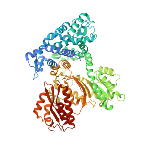
7UCH, 7UCI, 7UCL - PubMed Abstract:
Installation of methyl groups can significantly improve the binding of small-molecule drugs to protein targets; however, site-selective methylation often presents a significant synthetic challenge. Metal- and S -adenosyl-methionine (SAM)-dependent methyltransferases (MTs) in natural-product biosynthetic pathways are powerful enzymatic tools for selective or chemically challenging C-methylation reactions. Each of these MTs selectively catalyzes one or two methyl transfer reactions. Crystal structures and biochemical assays of the Mn 2+ -dependent monomethyltransferase from the saxitoxin biosynthetic pathway (SxtA MT) revealed the structural basis for control of methylation extent. The SxtA monomethyltransferase was converted to a dimethyltransferase by modification of the metal binding site, addition of an active site base, and an amino acid substitution to provide space in the substrate pocket for two methyl substituents. A reciprocal change converted a related dimethyltransferase into a monomethyltransferase, supporting our hypothesis that steric hindrance can prevent a second methylation event. A novel understanding of MTs will accelerate the development of MT-based catalysts and MT engineering for use in small-molecule synthesis.
- Program in Chemical Biology, University of Michigan, Ann Arbor, Michigan 48109, United States.
Organizational Affiliation: