Ir(III)-Based Agents for Monitoring the Cytochrome P450 3A4 Active Site Occupancy.
Denison, M., Steinke, S.J., Majeed, A., Turro, C., Kocarek, T.A., Sevrioukova, I.F., Kodanko, J.J.(2022) Inorg Chem 61: 13673-13677
- PubMed: 35994607 Search on PubMedSearch on PubMed Central
- DOI: https://doi.org/10.1021/acs.inorgchem.2c02587
- Primary Citation Related Structures:
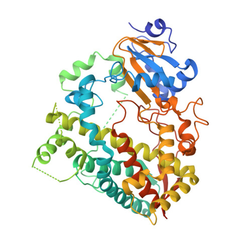
7UAY, 7UAZ - PubMed Abstract:
Cytochromes P450 (CYPs) are a superfamily of enzymes responsible for biosynthesis and drug metabolism. Monitoring the activity of CYP3A4, the major human drug-metabolizing enzyme, is vital for assessing the metabolism of pharmaceuticals and identifying harmful drug-drug interactions. Existing probes for CYP3A4 are irreversible turn-on substrates that monitor activity at specific time points in end-point assays. To provide a more dynamic approach, we designed, synthesized, and characterized emissive Ir(III) and Ru(II) complexes that allow monitoring of the CYP3A4 active-site occupancy in real time. In the bound state, probe emission is quenched by the active-site heme. Upon displacement from the active site by CYP3A4-specific inhibitors or substrates, these probes show high emission turn-on. Direct probe binding to the CYP3A4 active site was confirmed by X-ray crystallography. The lead Ir(III)-based probe has nanomolar K d and high selectivity for CYP3A4, efficient cellular uptake, and low toxicity in CYP3A4-overexpressing HepG2 cells.
- Department of Chemistry, Wayne State University, 5101 Cass Avenue, Detroit, Michigan 48202, United States.
Organizational Affiliation: