Ligand dependent interaction between PC-TP and PPAR delta mitigates diet-induced hepatic steatosis in male mice.
Druzak, S.A., Tardelli, M., Mays, S.G., El Bejjani, M., Mo, X., Maner-Smith, K.M., Bowen, T., Cato, M.L., Tillman, M.C., Sugiyama, A., Xie, Y., Fu, H., Cohen, D.E., Ortlund, E.A.(2023) Nat Commun 14: 2748-2748
- PubMed: 37173315 Search on PubMedSearch on PubMed Central
- DOI: https://doi.org/10.1038/s41467-023-38010-w
- Primary Citation Related Structures:
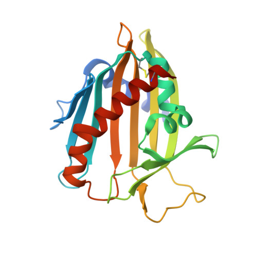
7U9D - PubMed Abstract:
Phosphatidylcholine transfer protein (PC-TP; synonym StarD2) is a soluble lipid-binding protein that transports phosphatidylcholine (PC) between cellular membranes. To better understand the protective metabolic effects associated with hepatic PC-TP, we generated a hepatocyte-specific PC-TP knockdown (L-Pctp -/- ) in male mice, which gains less weight and accumulates less liver fat compared to wild-type mice when challenged with a high-fat diet. Hepatic deletion of PC-TP also reduced adipose tissue mass and decreases levels of triglycerides and phospholipids in skeletal muscle, liver and plasma. Gene expression analysis suggest that the observed metabolic changes are related to transcriptional activity of peroxisome proliferative activating receptor (PPAR) family members. An in-cell protein complementation screen between lipid transfer proteins and PPARs uncovered a direct interaction between PC-TP and PPARδ that was not observed for other PPARs. We confirmed the PC-TP- PPARδ interaction in Huh7 hepatocytes, where it was found to repress PPARδ-mediated transactivation. Mutations of PC-TP residues implicated in PC binding and transfer reduce the PC-TP-PPARδ interaction and relieve PC-TP-mediated PPARδ repression. Reduction of exogenously supplied methionine and choline reduces the interaction while serum starvation enhances the interaction in cultured hepatocytes. Together our data points to a ligand sensitive PC-TP- PPARδ interaction that suppresses PPAR activity.
- Department of Biochemistry, Emory University School of Medicine, 1510 Clifton Road, Atlanta, GA, USA.
Organizational Affiliation: