7U8Z | pdb_00007u8z

Crystal structure of Danio rerio histone deacetylase 6 catalytic domain 2 (CD2) complexed with fluorinated peptoid inhibitor


Experimental Data Snapshot

  • Method: X-RAY DIFFRACTION
  • Resolution: 1.85 Å
  • R-Value Free: 
    0.215 (Depositor), 0.215 (DCC) 
  • R-Value Work: 
    0.172 (Depositor), 0.172 (DCC) 
  • R-Value Observed: 
    0.174 (Depositor) 

Starting Model: experimental
View more details

wwPDB Validation 3D Report Full Report

Validation slider image for 7U8Z

Ligand Structure Quality Assessment 


This is version 1.2 of the entry. See complete history

Literature

Development of Fluorinated Peptoid-Based Histone Deacetylase (HDAC) Inhibitors for Therapy-Resistant Acute Leukemia.

Ressing, N.Schliehe-Diecks, J.Watson, P.R.Sonnichsen, M.Cragin, A.D.Scholer, A.Yang, J.Schaker-Hubner, L.Borkhardt, A.Christianson, D.W.Bhatia, S.Hansen, F.K.

(2022) J Med Chem 65: 15457-15472

  • DOI: https://doi.org/10.1021/acs.jmedchem.2c01418
  • Primary Citation Related Structures: 
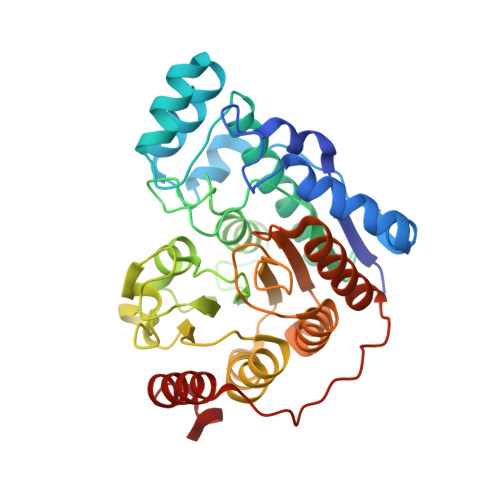
    7U8Z

  • PubMed Abstract: 

    Using a microwave-assisted protocol, we synthesized 16 peptoid-capped HDAC inhibitors (HDACi) with fluorinated linkers and identified two hit compounds. In biochemical and cellular assays, 10h stood out as a potent unselective HDACi with remarkable cytotoxic potential against different therapy-resistant leukemia cell lines. 10h demonstrated prominent antileukemic activity with low cytotoxic activity toward healthy cells. Moreover, 10h exhibited synergistic interactions with the DNA methyltransferase inhibitor decitabine in AML cell lines. The comparison of crystal structures of HDAC6 complexes with 10h and its nonfluorinated counterpart revealed a similar occupation of the L1 loop pocket but slight differences in zinc coordination. The substitution pattern of the acyl residue turned out to be crucial in terms of isoform selectivity. The introduction of an isopropyl group onto the phenyl ring provided the highly HDAC6-selective inhibitor 10p , which demonstrated moderate synergy with decitabine and exceeded the HDAC6 selectivity of tubastatin A.


  • Organizational Affiliation
    • Pharmaceutical Institute, Pharmaceutical and Cell Biological Chemistry, University of Bonn, An der Immenburg 4, 53121Bonn, Germany.

Macromolecule Content 

  • Total Structure Weight: 164.26 kDa 
  • Atom Count: 12,433 
  • Modeled Residue Count: 1,426 
  • Deposited Residue Count: 1,456 
  • Unique protein chains: 1

Macromolecules

Find similar proteins by:|  3D Structure
Entity ID: 1
MoleculeChains  Sequence LengthOrganismDetailsImage
Hdac6 protein
A, B, C, D
364Danio rerioMutation(s): 0 
Gene Names: hdac6
UniProt
Find proteins for F8W4B7 (Danio rerio)
Explore F8W4B7 
Go to UniProtKB:  F8W4B7
Entity Groups
Sequence Clusters30% Identity50% Identity70% Identity90% Identity95% Identity100% Identity
UniProt GroupF8W4B7
Sequence Annotations
Expand
Reference Sequence

Small Molecules

Ligands 7 Unique
IDChains Name / Formula / InChI Key2D Diagram3D Interactions
M0I

Query on M0I


Download:Ideal Coordinates CCD File
F [auth A],
J [auth B],
Q [auth C],
V [auth D]
4-({N-[2-(benzylamino)-2-oxoethyl]-4-(dimethylamino)benzamido}methyl)-3-fluoro-N-hydroxybenzamide
C26 H27 F N4 O4
GBIQEYYBFBGNOZ-UHFFFAOYSA-N
MLI

Query on MLI


Download:Ideal Coordinates CCD File
Z [auth D]MALONATE ION
C3 H2 O4
OFOBLEOULBTSOW-UHFFFAOYSA-L
GOL

Query on GOL


Download:Ideal Coordinates CCD File
R [auth C]GLYCEROL
C3 H8 O3
PEDCQBHIVMGVHV-UHFFFAOYSA-N
ZN

Query on ZN


Download:Ideal Coordinates CCD File
AA [auth D],
G [auth A],
N [auth B],
S [auth C]
ZINC ION
Zn
PTFCDOFLOPIGGS-UHFFFAOYSA-N
EDO

Query on EDO


Download:Ideal Coordinates CCD File
K [auth B]
L [auth B]
M [auth B]
W [auth D]
X [auth D]
K [auth B],
L [auth B],
M [auth B],
W [auth D],
X [auth D],
Y [auth D]
1,2-ETHANEDIOL
C2 H6 O2
LYCAIKOWRPUZTN-UHFFFAOYSA-N
ACT

Query on ACT


Download:Ideal Coordinates CCD File
E [auth A]ACETATE ION
C2 H3 O2
QTBSBXVTEAMEQO-UHFFFAOYSA-M
K

Query on K


Download:Ideal Coordinates CCD File
BA [auth D]
CA [auth D]
H [auth A]
I [auth A]
O [auth B]
BA [auth D],
CA [auth D],
H [auth A],
I [auth A],
O [auth B],
P [auth B],
T [auth C],
U [auth C]
POTASSIUM ION
K
NPYPAHLBTDXSSS-UHFFFAOYSA-N

Experimental Data & Validation

Experimental Data

  • Method: X-RAY DIFFRACTION
  • Resolution: 1.85 Å
  • R-Value Free:  0.215 (Depositor), 0.215 (DCC) 
  • R-Value Work:  0.172 (Depositor), 0.172 (DCC) 
  • R-Value Observed: 0.174 (Depositor) 
Space Group: P 1 21 1
Unit Cell:
Length ( Å )Angle ( ˚ )
a = 78.34α = 90
b = 95.13β = 98.73
c = 98.139γ = 90
Software Package:
Software NamePurpose
iMOSFLMdata reduction
PHENIXrefinement
Aimlessdata scaling
PHASERphasing

Structure Validation

View Full Validation Report


Ligand Structure Quality Assessment 


Entry History 

& Funding Information

Deposition Data


Funding OrganizationLocationGrant Number
National Institutes of Health/National Institute of General Medical Sciences (NIH/NIGMS)United StatesGM49758

Revision History  (Full details and data files)

  • Version 1.0: 2022-11-23
    Type: Initial release
  • Version 1.1: 2022-12-07
    Changes: Database references
  • Version 1.2: 2023-10-25
    Changes: Data collection, Refinement description