7U7H | pdb_00007u7h

Cysteate acyl-ACP transferase from Alistipes finegoldii


Experimental Data Snapshot

  • Method: X-RAY DIFFRACTION
  • Resolution: 2.90 Å
  • R-Value Free: 
    0.268 (Depositor), 0.246 (DCC) 
  • R-Value Work: 
    0.218 (Depositor), 0.223 (DCC) 
  • R-Value Observed: 
    0.221 (Depositor) 

Starting Model: experimental
View more details

wwPDB Validation 3D Report Full Report

Validation slider image for 7U7H

Ligand Structure Quality Assessment 


This is version 1.2 of the entry. See complete history

Literature

Biochemical characterization of the first step in sulfonolipid biosynthesis in Alistipes finegoldii.

Radka, C.D.Miller, D.J.Frank, M.W.Rock, C.O.

(2022) J Biological Chem 298: 102195-102195

  • DOI: https://doi.org/10.1016/j.jbc.2022.102195
  • Primary Citation Related Structures: 
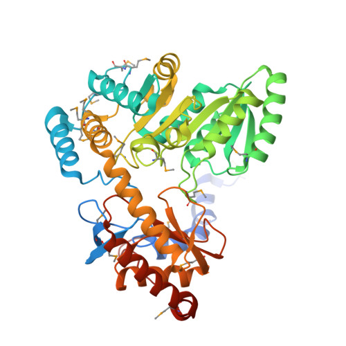
    7U7H

  • PubMed Abstract: 

    Sulfonolipids are unusual lipids found in the outer membranes of Gram-negative bacteria in the phylum Bacteroidetes. Sulfonolipid and its deacylated derivative, capnine, are sulfur analogs of ceramide-1-phosphate and sphingosine-1-phosphate, respectively; thus, sulfonolipid biosynthesis is postulated to be similar to the sphingolipid biosynthetic pathway. Here, we identify the first enzyme in sulfonolipid synthesis in Alistipes finegoldii as the product of the alfi_1224 gene, cysteate acyl-acyl carrier protein (ACP) transferase (SulA). We show SulA catalyzes the condensation of acyl-ACP and cysteate (3-sulfo-alanine) to form 3-ketocapnine. Acyl-CoA is a poor substrate. We show SulA has a bound pyridoxal phosphate (PLP) cofactor that undergoes a spectral redshift in the presence of cysteate, consistent with the transition of the lysine-aldimine complex to a substrate-aldimine complex. Furthermore, the SulA crystal structure shows the same prototypical fold found in bacterial serine palmitoyltransferases (Spts), enveloping the PLP cofactor bound to Lys251. We observed the SulA and Spt active sites are identical except for Lys281 in SulA, which is an alanine in Spt. Additionally, SulA(K281A) is catalytically inactive but binds cysteate and forms the external aldimine normally, highlighting the structural role of the Lys281 side chain in walling off the active site from bulk solvent. Finally, the electropositive groove on the protein surface adjacent to the active site entrance provides a landing pad for the electronegative acyl-ACP surface. Taken together, these data identify the substrates, products, and mechanism of SulA, the PLP-dependent condensing enzyme that catalyzes the first step in sulfonolipid synthesis in a gut commensal bacterium.


  • Organizational Affiliation
    • Department of Infectious Diseases, St. Jude Children's Research Hospital, Memphis, Tennessee, USA. Electronic address: christopher.radka@stjude.org.

Macromolecule Content 

  • Total Structure Weight: 300.71 kDa 
  • Atom Count: 18,499 
  • Modeled Residue Count: 2,492 
  • Deposited Residue Count: 2,658 
  • Unique protein chains: 1

Macromolecules

Find similar proteins by:|  3D Structure
Entity ID: 1
MoleculeChains  Sequence LengthOrganismDetailsImage
7-keto-8-aminopelargonate synthetase-like enzyme
A, B, C, D, E
A, B, C, D, E, F
443Alistipes finegoldii DSM 17242Mutation(s): 0 
Gene Names: Alfi_1224
UniProt
Find proteins for I3YKQ4 (Alistipes finegoldii (strain DSM 17242 / JCM 16770 / CCUG 46020 / CIP 107999 / KCTC 15236 / AHN 2437))
Explore I3YKQ4 
Go to UniProtKB:  I3YKQ4
Entity Groups
Sequence Clusters30% Identity50% Identity70% Identity90% Identity95% Identity100% Identity
UniProt GroupI3YKQ4
Sequence Annotations
Expand
Reference Sequence

Small Molecules

Modified Residues  1 Unique
IDChains TypeFormula2D DiagramParent
MSE
Query on MSE
A, B, C, D, E
A, B, C, D, E, F
L-PEPTIDE LINKINGC5 H11 N O2 SeMET

Experimental Data & Validation

Experimental Data

  • Method: X-RAY DIFFRACTION
  • Resolution: 2.90 Å
  • R-Value Free:  0.268 (Depositor), 0.246 (DCC) 
  • R-Value Work:  0.218 (Depositor), 0.223 (DCC) 
  • R-Value Observed: 0.221 (Depositor) 
Space Group: P 31
Unit Cell:
Length ( Å )Angle ( ˚ )
a = 127.516α = 90
b = 127.516β = 90
c = 177.3γ = 120
Software Package:
Software NamePurpose
PHENIXrefinement
PDB_EXTRACTdata extraction
XDSdata reduction
XDSdata scaling
PHENIXphasing

Structure Validation

View Full Validation Report


Ligand Structure Quality Assessment 


Entry History 

& Funding Information

Deposition Data


Funding OrganizationLocationGrant Number
National Institutes of Health/National Institute Of Allergy and Infectious Diseases (NIH/NIAID)United States1K99AI166116-01
National Institutes of Health/National Institute of General Medical Sciences (NIH/NIGMS)United States5R01GM034496-38

Revision History  (Full details and data files)

  • Version 1.0: 2023-01-18
    Type: Initial release
  • Version 1.1: 2023-10-25
    Changes: Data collection, Refinement description
  • Version 1.2: 2023-11-15
    Changes: Data collection